4FXE

Crystal structure of the intact E. coli RelBE toxin-antitoxin complex


Experimental Data Snapshot

  • Method: X-RAY DIFFRACTION
  • Resolution: 2.75 Å
  • R-Value Free: 0.285 
  • R-Value Work: 0.253 
  • R-Value Observed: 0.255 

wwPDB Validation   3D Report Full Report


This is version 1.3 of the entry. See complete history


Literature

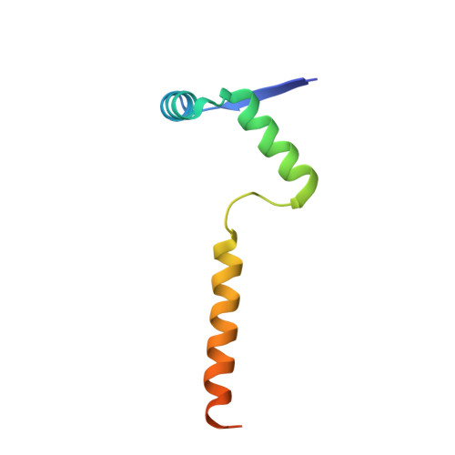
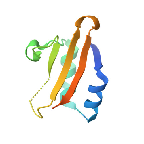
The crystal structure of the intact E. coli RelBE toxin-antitoxin complex provides the structural basis for conditional cooperativity.

Boggild, A.Sofos, N.Andersen, K.R.Feddersen, A.Easter, A.D.Passmore, L.A.Brodersen, D.E.

(2012) Structure 20: 1641-1648

  • DOI: https://doi.org/10.1016/j.str.2012.08.017
  • Primary Citation of Related Structures:  
    4FXE, 4FXH, 4FXI

  • PubMed Abstract: 

    The bacterial relBE locus encodes a toxin-antitoxin complex in which the toxin, RelE, is capable of cleaving mRNA in the ribosomal A site cotranslationally. The antitoxin, RelB, both binds and inhibits RelE, and regulates transcription through operator binding and conditional cooperativity controlled by RelE. Here, we present the crystal structure of the intact Escherichia coli RelB2E2 complex at 2.8 Å resolution, comprising both the RelB-inhibited RelE and the RelB dimerization domain that binds DNA. RelE and RelB associate into a V-shaped heterotetrameric complex with the ribbon-helix-helix (RHH) dimerization domain at the apex. Our structure supports a model in which relO is optimally bound by two adjacent RelB2E heterotrimeric units, and is not compatible with concomitant binding of two RelB2E2 heterotetramers. The results thus provide a firm basis for understanding the model of conditional cooperativity at the molecular level.


  • Organizational Affiliation

    Centre for mRNP Biogenesis and Metabolism, Department of Molecular Biology and Genetics, Aarhus University, DK-8000 Aarhus C, Denmark.


Macromolecules
Find similar proteins by:  (by identity cutoff)  |  3D Structure
Entity ID: 1
MoleculeChains Sequence LengthOrganismDetailsImage
Antitoxin RelB
A, B, C
79Escherichia coli K-12Mutation(s): 0 
Gene Names: b1564JW1556relB
UniProt
Find proteins for P0C079 (Escherichia coli (strain K12))
Explore P0C079 
Go to UniProtKB:  P0C079
Entity Groups  
Sequence Clusters30% Identity50% Identity70% Identity90% Identity95% Identity100% Identity
UniProt GroupP0C079
Sequence Annotations
Expand
  • Reference Sequence
Find similar proteins by:  (by identity cutoff)  |  3D Structure
Entity ID: 2
MoleculeChains Sequence LengthOrganismDetailsImage
mRNA interferase RelE
D, E, F
95Escherichia coli K-12Mutation(s): 1 
Gene Names: b1563JW1555relE
EC: 3.1
UniProt
Find proteins for P0C077 (Escherichia coli (strain K12))
Explore P0C077 
Go to UniProtKB:  P0C077
Entity Groups  
Sequence Clusters30% Identity50% Identity70% Identity90% Identity95% Identity100% Identity
UniProt GroupP0C077
Sequence Annotations
Expand
  • Reference Sequence
Experimental Data & Validation

Experimental Data

  • Method: X-RAY DIFFRACTION
  • Resolution: 2.75 Å
  • R-Value Free: 0.285 
  • R-Value Work: 0.253 
  • R-Value Observed: 0.255 
  • Space Group: P 61 2 2
Unit Cell:
Length ( Å )Angle ( ˚ )
a = 77.23α = 90
b = 77.23β = 90
c = 362.82γ = 120
Software Package:
Software NamePurpose
XSCALEdata scaling
SHARPphasing
SOLOMONphasing
PHENIXrefinement
PDB_EXTRACTdata extraction
XDSdata reduction

Structure Validation

View Full Validation Report


Entry History 

Deposition Data

Revision History  (Full details and data files)

  • Version 1.0: 2012-08-29
    Type: Initial release
  • Version 1.1: 2012-12-26
    Changes: Database references
  • Version 1.2: 2018-03-07
    Changes: Data collection
  • Version 1.3: 2024-02-28
    Changes: Data collection, Database references, Derived calculations