


Crystal structure of a probable oxidoreduxtase protein
Eswaramoorthy, S., Chamala, S., Evans, B., Foti, R., Gizzi, A., Hillerich, B., Kar, A., Lafleur, J., Seidel, R., Villigas, G., Zencheck, W., Almo, S.C., Swaminathan, S.To be published.
Experimental Data Snapshot
wwPDB Validation 3D Report Full Report

Entity ID: 1 | |||||
|---|---|---|---|---|---|
| Molecule | Chains | Sequence Length | Organism | Details | Image |
| Probable oxidoreductase protein | 393 | Rhizobium etli CFN 42 | Mutation(s): 0 Gene Names: RHE_CH01346 | 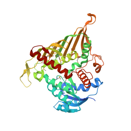 | |
UniProt | |||||
Find proteins for Q2KAI6 (Rhizobium etli (strain CFN 42 / ATCC 51251)) Explore Q2KAI6 Go to UniProtKB: Q2KAI6 | |||||
Entity Groups | |||||
| Sequence Clusters | 30% Identity50% Identity70% Identity90% Identity95% Identity100% Identity | ||||
| UniProt Group | Q2KAI6 | ||||
Sequence AnnotationsExpand | |||||
| |||||
| Modified Residues 1 Unique | |||||
|---|---|---|---|---|---|
| ID | Chains | Type | Formula | 2D Diagram | Parent |
| MSE Query on MSE | A, B | L-PEPTIDE LINKING | C5 H11 N O2 Se |  | MET |
| Length ( Å ) | Angle ( ˚ ) |
|---|---|
| a = 87.342 | α = 90 |
| b = 179.094 | β = 90 |
| c = 127.824 | γ = 90 |
| Software Name | Purpose |
|---|---|
| CBASS | data collection |
| SHELXS | phasing |
| REFMAC | refinement |
| HKL-2000 | data reduction |
| HKL-2000 | data scaling |
RCSB PDB (citation) is hosted by
RCSB PDB is a member of the

