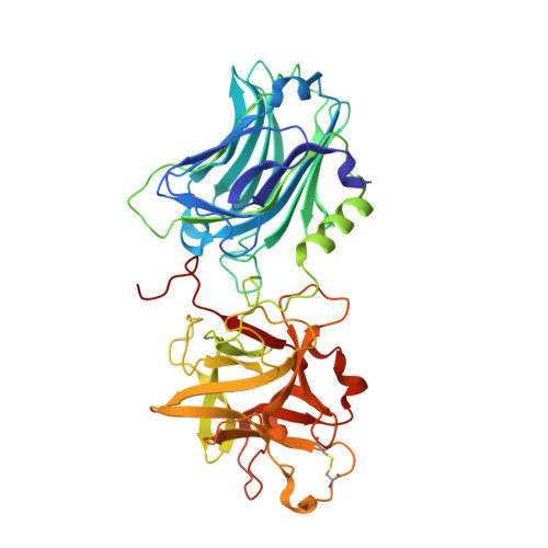
Structural insights into the functional role of the Hcn sub-domain of the receptor-binding domain of the botulinum neurotoxin mosaic serotype C/D.
Zhang, Y., Gardberg, A.S., Edwards, T.E., Sankaran, B., Robinson, H., Varnum, S.M., Buchko, G.W.(2013) Biochimie 95: 1379-1385
- PubMed: 23523511
- DOI: https://doi.org/10.1016/j.biochi.2013.03.006
- Primary Citation of Related Structures:
4F83 - PubMed Abstract:
Botulinum neurotoxin (BoNT), the causative agent of the deadly neuroparalytic disease botulism, is the most poisonous protein known for humans. Produced by different strains of the anaerobic bacterium Clostridium botulinum, BoNT effects cellular intoxication via a multistep mechanism executed by the three modules of the activated protein. Endocytosis, the first step of cellular intoxication, is triggered by the ~50 kDa, heavy-chain receptor-binding domain (HCR) that is specific for a ganglioside and a protein receptor on neuronal cell surfaces. This dual receptor recognition mechanism between BoNT and the host cell's membrane is well documented and occurs via specific intermolecular interactions with the C-terminal sub-domain, Hcc, of BoNT-HCR. The N-terminal sub-domain of BoNT-HCR, Hcn, comprises ~50% of BoNT-HCR and adopts a β-sheet jelly roll fold. While suspected in assisting cell surface recognition, no unambiguous function for the Hcn sub-domain in BoNT has been identified. To obtain insights into the potential function of the Hcn sub-domain in BoNT, the first crystal structure of a BoNT with an organic ligand bound to the Hcn sub-domain has been obtained. Here, we describe the crystal structure of BoNT/CD-HCR determined at 1.70 Å resolution with a tetraethylene glycol (PG4) moiety bound in a hydrophobic cleft between β-strands in the β-sheet jelly roll fold of the Hcn sub-domain. The PG4 moiety is completely engulfed in the cleft, making numerous hydrophilic (Y932, S959, W966, and D1042) and hydrophobic (S935, W977, L979, N1013, and I1066) contacts with the protein's side chain and backbone that may mimic in vivo interactions with the phospholipid membranes on neuronal cell surfaces. A sulfate ion was also observed bound to residues T1176, D1177, K1196, and R1243 in the Hcc sub-domain of BoNT/CD-HCR. In the crystal structure of a similar protein, BoNT/D-HCR, a sialic acid molecule was observed bound to the equivalent residues suggesting that residues T1176, D1177, K1196, and R1243 in BoNT/CD may play a role in ganglioside binding.
Organizational Affiliation:
Biological Sciences Division, Pacific Northwest National Laboratory, Richland, WA 99352, USA.


