


Crystal structure of Nucleoside diphosphate kinase B from Trypanosoma brucei, apo form
Seattle Structural Genomics Center for Infectious Disease (SSGCID), Gardberg, A.S., Edwards, T.E., Staker, B., Stewart, L.To be published.
Experimental Data Snapshot
wwPDB Validation 3D Report Full Report

Entity ID: 1 | |||||
|---|---|---|---|---|---|
| Molecule | Chains | Sequence Length | Organism | Details | Image |
| Nucleoside diphosphate kinase | 157 | Trypanosoma brucei brucei TREU927 | Mutation(s): 0 Gene Names: Tb11.01.7800 EC: 2.7.4.6 | 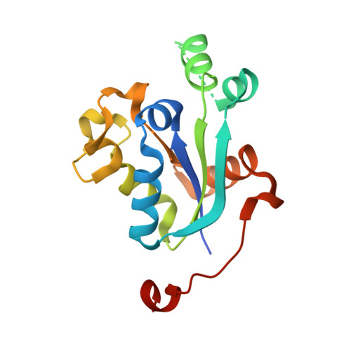 | |
UniProt | |||||
Find proteins for Q381H3 (Trypanosoma brucei brucei (strain 927/4 GUTat10.1)) Explore Q381H3 Go to UniProtKB: Q381H3 | |||||
Entity Groups | |||||
| Sequence Clusters | 30% Identity50% Identity70% Identity90% Identity95% Identity100% Identity | ||||
| UniProt Group | Q381H3 | ||||
Sequence AnnotationsExpand | |||||
| |||||
| Ligands 1 Unique | |||||
|---|---|---|---|---|---|
| ID | Chains | Name / Formula / InChI Key | 2D Diagram | 3D Interactions | |
| SCN Query on SCN | G [auth F] | THIOCYANATE ION C N S ZMZDMBWJUHKJPS-UHFFFAOYSA-M |  | ||
| Length ( Å ) | Angle ( ˚ ) |
|---|---|
| a = 52.46 | α = 90 |
| b = 123.67 | β = 90 |
| c = 145.36 | γ = 90 |
| Software Name | Purpose |
|---|---|
| XSCALE | data scaling |
| PHASER | phasing |
| REFMAC | refinement |
| PDB_EXTRACT | data extraction |
| XDS | data reduction |
RCSB PDB (citation) is hosted by
RCSB PDB is a member of the

