4ENH

Crystal Structure of Human Cytochrome P450 CYP46A1 with Fluvoxamine Bound


Experimental Data Snapshot

  • Method: X-RAY DIFFRACTION
  • Resolution: 2.50 Å
  • R-Value Free: 0.272 
  • R-Value Work: 0.210 
  • R-Value Observed: 0.213 

wwPDB Validation   3D Report Full Report


Ligand Structure Quality Assessment 


This is version 1.2 of the entry. See complete history


Literature

In Silico and Intuitive Predictions of CYP46A1 Inhibition by Marketed Drugs with Subsequent Enzyme Crystallization in Complex with Fluvoxamine.

Mast, N.Linger, M.Clark, M.Wiseman, J.Stout, C.D.Pikuleva, I.A.

(2012) Mol Pharmacol 82: 824-834

  • DOI: https://doi.org/10.1124/mol.112.080424
  • Primary Citation of Related Structures:  
    4ENH

  • PubMed Abstract: 

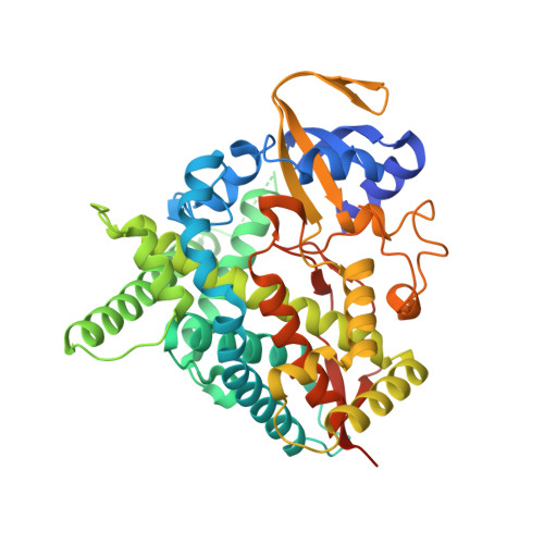
    Cytochrome P450 46A1 (cholesterol 24-hydroxylase) is an important brain enzyme that may be inhibited by structurally distinct pharmaceutical agents both in vitro and in vivo. To identify additional inhibitors of CYP46A1 among U.S. Food and Drug Administration-approved therapeutic agents, we used in silico and intuitive predictions and evaluated some of the predicted binders in the enzyme and spectral binding assays. We tested a total of 298 marketed drugs for the inhibition of CYP46A1-mediated cholesterol hydroxylation in vitro and found that 13 of them reduce CYP46A1 activity by >50%. Of these 13 inhibitors, 7 elicited a spectral response in CYP46A1 with apparent spectral K(d) values in a low micromolar range. One of the identified tight binders, the widely used antidepressant fluvoxamine, was cocrystallized with CYP46A1. The structure of this complex was determined at a 2.5 Å resolution and revealed the details of drug binding to the CYP46A1 active site. The NH(2)-containing arm of the Y-shaped fluvoxamine coordinates the CYP46A1 heme iron, whereas the methoxy-containing arm points away from the heme group and has multiple hydrophobic interactions with aliphatic amino acid residues. The CF(3)-phenyl ring faces the entrance to the substrate access channel and has contacts with the aromatic side chains. The crystal structure suggests that only certain drug conformers can enter the P450 substrate access channel and reach the active site. Once inside the active site, the conformer probably further adjusts its configuration and elicits the movement of the protein side chains.


  • Organizational Affiliation

    Department of Ophthalmology and Visual Sciences, Case Western Reserve University, Cleveland, Ohio 44106, USA.


Macromolecules
Find similar proteins by:  (by identity cutoff)  |  3D Structure
Entity ID: 1
MoleculeChains Sequence LengthOrganismDetailsImage
Cholesterol 24-hydroxylase456Homo sapiensMutation(s): 0 
Gene Names: CYP46A1CYP46
EC: 1.14.13.98
UniProt & NIH Common Fund Data Resources
Find proteins for Q9Y6A2 (Homo sapiens)
Explore Q9Y6A2 
Go to UniProtKB:  Q9Y6A2
PHAROS:  Q9Y6A2
GTEx:  ENSG00000036530 
Entity Groups  
Sequence Clusters30% Identity50% Identity70% Identity90% Identity95% Identity100% Identity
UniProt GroupQ9Y6A2
Sequence Annotations
Expand
  • Reference Sequence
Small Molecules
Ligands 2 Unique
IDChains Name / Formula / InChI Key2D Diagram3D Interactions
HEM
Query on HEM

Download Ideal Coordinates CCD File 
B [auth A]PROTOPORPHYRIN IX CONTAINING FE
C34 H32 Fe N4 O4
KABFMIBPWCXCRK-RGGAHWMASA-L
FVX
Query on FVX

Download Ideal Coordinates CCD File 
C [auth A]Fluvoxamine
C15 H21 F3 N2 O2
CJOFXWAVKWHTFT-XSFVSMFZSA-N
Binding Affinity Annotations 
IDSourceBinding Affinity
FVX Binding MOAD:  4ENH Ki: 1700 (nM) from 1 assay(s)
Experimental Data & Validation

Experimental Data

  • Method: X-RAY DIFFRACTION
  • Resolution: 2.50 Å
  • R-Value Free: 0.272 
  • R-Value Work: 0.210 
  • R-Value Observed: 0.213 
  • Space Group: I 41 2 2
Unit Cell:
Length ( Å )Angle ( ˚ )
a = 121.13α = 90
b = 121.13β = 90
c = 141.9γ = 90
Software Package:
Software NamePurpose
SCALAdata scaling
REFMACrefinement
PDB_EXTRACTdata extraction
Blu-Icedata collection
XDSdata reduction
PHASERphasing

Structure Validation

View Full Validation Report


Ligand Structure Quality Assessment 


Entry History 

Deposition Data

Revision History  (Full details and data files)

  • Version 1.0: 2012-08-22
    Type: Initial release
  • Version 1.1: 2012-12-19
    Changes: Database references
  • Version 1.2: 2023-09-13
    Changes: Data collection, Database references, Derived calculations, Refinement description