


Crystal structure of aminotransferase from anaerococcus prevotii dsm 20548.
Chang, C., Tesar, C., Bearden, J., Joachimiak, A.To be published.
Experimental Data Snapshot
Entity ID: 1 | |||||
|---|---|---|---|---|---|
| Molecule | Chains | Sequence Length | Organism | Details | Image |
| Aminotransferase class I and II | 413 | Anaerococcus prevotii DSM 20548 | Mutation(s): 0 Gene Names: Apre_1502 | 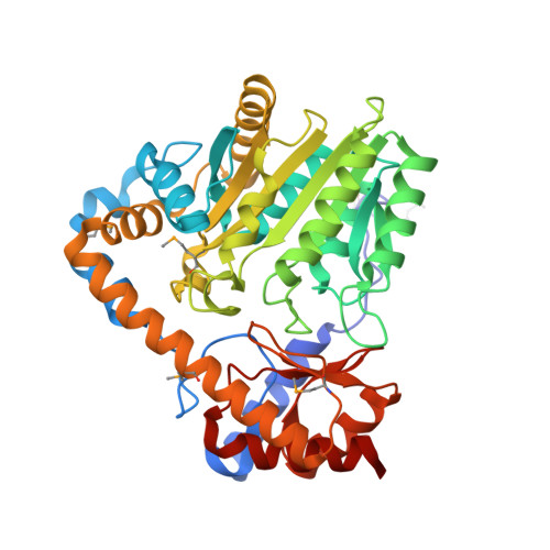 | |
UniProt | |||||
Find proteins for C7REB0 (Anaerococcus prevotii (strain ATCC 9321 / DSM 20548 / JCM 6508 / NCTC 11806 / PC1)) Explore C7REB0 Go to UniProtKB: C7REB0 | |||||
Entity Groups | |||||
| Sequence Clusters | 30% Identity50% Identity70% Identity90% Identity95% Identity100% Identity | ||||
| UniProt Group | C7REB0 | ||||
Sequence AnnotationsExpand | |||||
| |||||
| Ligands 1 Unique | |||||
|---|---|---|---|---|---|
| ID | Chains | Name / Formula / InChI Key | 2D Diagram | 3D Interactions | |
| PLP Query on PLP | E [auth A], F [auth B], G [auth C], H [auth D] | PYRIDOXAL-5'-PHOSPHATE C8 H10 N O6 P NGVDGCNFYWLIFO-UHFFFAOYSA-N |  | ||
| Modified Residues 1 Unique | |||||
|---|---|---|---|---|---|
| ID | Chains | Type | Formula | 2D Diagram | Parent |
| MSE Query on MSE | A, B, C, D | L-PEPTIDE LINKING | C5 H11 N O2 Se |  | MET |
| Length ( Å ) | Angle ( ˚ ) |
|---|---|
| a = 64.297 | α = 90 |
| b = 131.178 | β = 90 |
| c = 239.913 | γ = 90 |
| Software Name | Purpose |
|---|---|
| REFMAC | refinement |
| PDB_EXTRACT | data extraction |
| HKL-3000 | data reduction |
| HKL-3000 | data scaling |
| HKL-3000 | phasing |
| MLPHARE | phasing |
| DM | phasing |
| SHELXDE | phasing |
| RESOLVE | phasing |
| ARP/wARP | model building |
| Coot | model building |
RCSB PDB (citation) is hosted by
RCSB PDB is a member of the

