


The Structure of Ribose 5-phosphate Isomerase B from Anaplasma phagocytophilum
Clifton, M.C., Edwards, T.E., Sankaran, B., Seattle Structural Genomics Center for Infectious Disease (SSGCID)To be published.
Experimental Data Snapshot
wwPDB Validation 3D Report Full Report

Entity ID: 1 | |||||
|---|---|---|---|---|---|
| Molecule | Chains | Sequence Length | Organism | Details | Image |
| Ribose 5-phosphate isomerase B | 148 | Anaplasma phagocytophilum str. HZ | Mutation(s): 0 Gene Names: rpiB, APH_0642 EC: 5.3.1.6 | 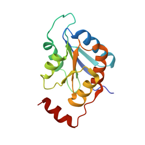 | |
UniProt | |||||
Find proteins for Q2GK74 (Anaplasma phagocytophilum (strain HZ)) Explore Q2GK74 Go to UniProtKB: Q2GK74 | |||||
Entity Groups | |||||
| Sequence Clusters | 30% Identity50% Identity70% Identity90% Identity95% Identity100% Identity | ||||
| UniProt Group | Q2GK74 | ||||
Sequence AnnotationsExpand | |||||
| |||||
| Length ( Å ) | Angle ( ˚ ) |
|---|---|
| a = 35.36 | α = 90 |
| b = 62.49 | β = 93.13 |
| c = 73.96 | γ = 90 |
| Software Name | Purpose |
|---|---|
| XSCALE | data scaling |
| PHASER | phasing |
| REFMAC | refinement |
| PDB_EXTRACT | data extraction |
| XDS | data reduction |
RCSB PDB (citation) is hosted by
RCSB PDB is a member of the

