4E89

Crystal Structure of RnaseH from gammaretrovirus


Experimental Data Snapshot

  • Method: X-RAY DIFFRACTION
  • Resolution: 2.60 Å
  • R-Value Free: 0.260 
  • R-Value Work: 0.239 
  • R-Value Observed: 0.239 

wwPDB Validation   3D Report Full Report


This is version 1.1 of the entry. See complete history


Literature

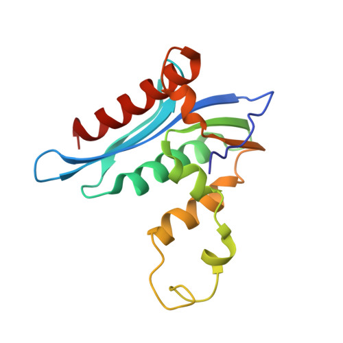
Crystal structure of xenotropic murine leukaemia virus-related virus (XMRV) ribonuclease H

Kim, J.H.Kang, S.Jung, S.K.Yu, K.R.Chung, S.J.Chung, B.H.Erikson, R.L.Kim, B.Y.Kim, S.J.

(2012) Biosci Rep 32: 455-463

  • DOI: https://doi.org/10.1042/BSR20120028
  • Primary Citation of Related Structures:  
    4E89

  • PubMed Abstract: 

    RNase H (retroviral ribonuclease H) cleaves the phosphate backbone of the RNA template within an RNA/DNA hybrid to complete the synthesis of double-stranded viral DNA. In the present study we have determined the complete structure of the RNase H domain from XMRV (xenotropic murine leukaemia virus-related virus) RT (reverse transcriptase). The basic protrusion motif of the XMRV RNase H domain is folded as a short helix and an adjacent highly bent loop. Structural superposition and subsequent mutagenesis experiments suggest that the basic protrusion motif plays a role in direct binding to the major groove in RNA/DNA hybrid, as well as in establishing the co-ordination among modules in RT necessary for proper function.


  • Organizational Affiliation

    Medical Proteomics Research Center, Korea Research Institute of Bioscience and Biotechnology, Yuseong-Gu, Daejeon, Republic of Korea.


Macromolecules
Find similar proteins by:  (by identity cutoff)  |  3D Structure
Entity ID: 1
MoleculeChains Sequence LengthOrganismDetailsImage
RNase H157Xenotropic MuLV-related virus VP62Mutation(s): 0 
EC: 3.1.26.4
UniProt
Find proteins for A1Z651 (Xenotropic MuLV-related virus (isolate VP62))
Explore A1Z651 
Go to UniProtKB:  A1Z651
Entity Groups  
Sequence Clusters30% Identity50% Identity70% Identity90% Identity95% Identity100% Identity
UniProt GroupA1Z651
Sequence Annotations
Expand
  • Reference Sequence
Experimental Data & Validation

Experimental Data

  • Method: X-RAY DIFFRACTION
  • Resolution: 2.60 Å
  • R-Value Free: 0.260 
  • R-Value Work: 0.239 
  • R-Value Observed: 0.239 
  • Space Group: P 43 21 2
Unit Cell:
Length ( Å )Angle ( ˚ )
a = 63.7α = 90
b = 63.7β = 90
c = 163.92γ = 90
Software Package:
Software NamePurpose
ADSCdata collection
PHASERphasing
CNSrefinement
MOSFLMdata reduction
SCALAdata scaling

Structure Validation

View Full Validation Report


Entry History 

Deposition Data

Revision History  (Full details and data files)

  • Version 1.0: 2012-10-17
    Type: Initial release
  • Version 1.1: 2024-03-20
    Changes: Data collection, Database references, Derived calculations