4E6K

2.0 A resolution structure of Pseudomonas aeruginosa bacterioferritin (BfrB) in complex with bacterioferritin associated ferredoxin (Bfd)


Experimental Data Snapshot

  • Method: X-RAY DIFFRACTION
  • Resolution: 2.00 Å
  • R-Value Free: 0.183 
  • R-Value Work: 0.152 
  • R-Value Observed: 0.154 

wwPDB Validation   3D Report Full Report


Ligand Structure Quality Assessment 


This is version 1.3 of the entry. See complete history


Literature

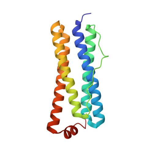
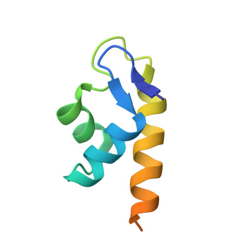
The Structure of the BfrB-Bfd Complex Reveals Protein-Protein Interactions Enabling Iron Release from Bacterioferritin.

Yao, H.Wang, Y.Lovell, S.Kumar, R.Ruvinsky, A.M.Battaile, K.P.Vakser, I.A.Rivera, M.

(2012) J Am Chem Soc 134: 13470-13481

  • DOI: https://doi.org/10.1021/ja305180n
  • Primary Citation of Related Structures:  
    4E6K

  • PubMed Abstract: 

    Ferritin-like molecules are unique to cellular iron homeostasis because they can store iron at concentrations much higher than those dictated by the solubility of Fe(3+). Very little is known about the protein interactions that deliver iron for storage or promote the mobilization of stored iron from ferritin-like molecules. Here, we report the X-ray crystal structure of Pseudomonas aeruginosa bacterioferritin (Pa-BfrB) in complex with bacterioferritin-associated ferredoxin (Pa-Bfd) at 2.0 Å resolution. As the first example of a ferritin-like molecule in complex with a cognate partner, the structure provides unprecedented insight into the complementary interface that enables the [2Fe-2S] cluster of Pa-Bfd to promote heme-mediated electron transfer through the BfrB protein dielectric (~18 Å), a process that is necessary to reduce the core ferric mineral and facilitate mobilization of Fe(2+). The Pa-BfrB-Bfd complex also revealed the first structure of a Bfd, thus providing a first view to what appears to be a versatile metal binding domain ubiquitous to the large Fer2_BFD family of proteins and enzymes with diverse functions. Residues at the Pa-BfrB-Bfd interface are highly conserved in Bfr and Bfd sequences from a number of pathogenic bacteria, suggesting that the specific recognition between Pa-BfrB and Pa-Bfd is of widespread significance to the understanding of bacterial iron homeostasis.


  • Organizational Affiliation

    Department of Chemistry, University of Kansas, Multidisciplinary Research Building, 2030 Becker Drive, Lawrence, Kansas 66047, USA.


Macromolecules
Find similar proteins by:  (by identity cutoff)  |  3D Structure
Entity ID: 1
MoleculeChains Sequence LengthOrganismDetailsImage
Bacterioferritin
A, B, C, D, E
A, B, C, D, E, F
158Pseudomonas aeruginosa PAO1Mutation(s): 0 
Gene Names: bfrBPA3531
UniProt
Find proteins for Q9HY79 (Pseudomonas aeruginosa (strain ATCC 15692 / DSM 22644 / CIP 104116 / JCM 14847 / LMG 12228 / 1C / PRS 101 / PAO1))
Explore Q9HY79 
Go to UniProtKB:  Q9HY79
Entity Groups  
Sequence Clusters30% Identity50% Identity70% Identity90% Identity95% Identity100% Identity
UniProt GroupQ9HY79
Sequence Annotations
Expand
  • Reference Sequence
Find similar proteins by:  (by identity cutoff)  |  3D Structure
Entity ID: 2
MoleculeChains Sequence LengthOrganismDetailsImage
bacterioferritin-associated ferredoxin
G, H, I
73Pseudomonas aeruginosa PAO1Mutation(s): 1 
Gene Names: BFDPA3530
UniProt
Find proteins for Q9HY80 (Pseudomonas aeruginosa (strain ATCC 15692 / DSM 22644 / CIP 104116 / JCM 14847 / LMG 12228 / 1C / PRS 101 / PAO1))
Explore Q9HY80 
Go to UniProtKB:  Q9HY80
Entity Groups  
Sequence Clusters30% Identity50% Identity70% Identity90% Identity95% Identity100% Identity
UniProt GroupQ9HY80
Sequence Annotations
Expand
  • Reference Sequence
Small Molecules
Ligands 5 Unique
IDChains Name / Formula / InChI Key2D Diagram3D Interactions
HEM
Query on HEM

Download Ideal Coordinates CCD File 
K [auth A],
O [auth C],
R [auth E]
PROTOPORPHYRIN IX CONTAINING FE
C34 H32 Fe N4 O4
KABFMIBPWCXCRK-RGGAHWMASA-L
FES
Query on FES

Download Ideal Coordinates CCD File 
V [auth G],
X [auth H],
Y [auth I]
FE2/S2 (INORGANIC) CLUSTER
Fe2 S2
NIXDOXVAJZFRNF-UHFFFAOYSA-N
PO4
Query on PO4

Download Ideal Coordinates CCD File 
W [auth G]PHOSPHATE ION
O4 P
NBIIXXVUZAFLBC-UHFFFAOYSA-K
K
Query on K

Download Ideal Coordinates CCD File 
J [auth A],
M [auth B],
T [auth F]
POTASSIUM ION
K
NPYPAHLBTDXSSS-UHFFFAOYSA-N
NA
Query on NA

Download Ideal Coordinates CCD File 
L [auth A]
N [auth B]
P [auth C]
Q [auth D]
S [auth E]
L [auth A],
N [auth B],
P [auth C],
Q [auth D],
S [auth E],
U [auth F]
SODIUM ION
Na
FKNQFGJONOIPTF-UHFFFAOYSA-N
Experimental Data & Validation

Experimental Data

  • Method: X-RAY DIFFRACTION
  • Resolution: 2.00 Å
  • R-Value Free: 0.183 
  • R-Value Work: 0.152 
  • R-Value Observed: 0.154 
  • Space Group: P 4 21 2
Unit Cell:
Length ( Å )Angle ( ˚ )
a = 135.005α = 90
b = 135.005β = 90
c = 200.89γ = 90
Software Package:
Software NamePurpose
SCALAdata scaling
PHASERphasing
PHENIXrefinement
PDB_EXTRACTdata extraction
JDirectordata collection
XDSdata reduction

Structure Validation

View Full Validation Report


Ligand Structure Quality Assessment 


Entry History 

Deposition Data

Revision History  (Full details and data files)

  • Version 1.0: 2012-08-01
    Type: Initial release
  • Version 1.1: 2012-08-29
    Changes: Database references
  • Version 1.2: 2017-11-15
    Changes: Refinement description
  • Version 1.3: 2023-09-13
    Changes: Data collection, Database references, Derived calculations, Refinement description