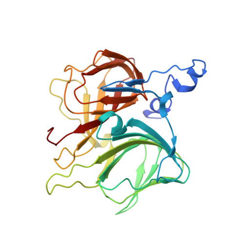
4E2Q

Crystal Structure of (S)-Ureidoglycine Aminohydrolase from Arabidopsis thaliana


Experimental Data Snapshot

  • Method: X-RAY DIFFRACTION
  • Resolution: 2.50 Å
  • R-Value Free: 0.286 
  • R-Value Work: 0.224 

wwPDB Validation   3D Report Full Report


This is version 1.2 of the entry. See complete history


Literature

Structural and functional insights into (S)-ureidoglycine aminohydrolase, key enzyme of purine catabolism in Arabidopsis thaliana

Shin, I.Percudani, R.Rhee, S.

(2012) J Biol Chem 287: 18796-18805

  • DOI: https://doi.org/10.1074/jbc.M111.331819
  • Primary Citation of Related Structures:  
    4E2Q, 4E2S

  • PubMed Abstract: 

    The ureide pathway has recently been identified as the metabolic route of purine catabolism in plants and some bacteria. In this pathway, uric acid, which is a major product of the early stage of purine catabolism, is degraded into glyoxylate and ammonia via stepwise reactions of seven different enzymes. Therefore, the pathway has a possible physiological role in mobilization of purine ring nitrogen for further assimilation. (S)-Ureidoglycine aminohydrolase enzyme converts (S)-ureidoglycine into (S)-ureidoglycolate and ammonia, providing the final substrate to the pathway. Here, we report a structural and functional analysis of this enzyme from Arabidopsis thaliana (AtUGlyAH). The crystal structure of AtUGlyAH in the ligand-free form shows a monomer structure in the bicupin fold of the β-barrel and an octameric functional unit as well as a Mn(2+) ion binding site. The structure of AtUGlyAH in complex with (S)-ureidoglycine revealed that the Mn(2+) ion acts as a molecular anchor to bind (S)-ureidoglycine, and its binding mode dictates the enantioselectivity of the reaction. Further kinetic analysis characterized the functional roles of the active site residues, including the Mn(2+) ion binding site and residues in the vicinity of (S)-ureidoglycine. These analyses provide molecular insights into the structure of the enzyme and its possible catalytic mechanism.


  • Organizational Affiliation

    Department of Agricultural Biotechnology, Seoul National University, Seoul, Korea.


Macromolecules
Find similar proteins by:  (by identity cutoff)  |  3D Structure
Entity ID: 1
MoleculeChains Sequence LengthOrganismDetailsImage
Ureidoglycine aminohydrolase
A, B, C, D, E
A, B, C, D, E, F, G, H, I, J, K, L, M, N, O, P
266Arabidopsis thalianaMutation(s): 0 
Gene Names: ylbAUGLYAHAt4g17050
EC: 3.5.3
UniProt
Find proteins for Q8GXV5 (Arabidopsis thaliana)
Explore Q8GXV5 
Go to UniProtKB:  Q8GXV5
Entity Groups  
Sequence Clusters30% Identity50% Identity70% Identity90% Identity95% Identity100% Identity
UniProt GroupQ8GXV5
Sequence Annotations
Expand
  • Reference Sequence
Small Molecules
Ligands 1 Unique
IDChains Name / Formula / InChI Key2D Diagram3D Interactions
MN
Query on MN

Download Ideal Coordinates CCD File 
AA [auth K]
BA [auth L]
CA [auth M]
DA [auth N]
EA [auth O]
AA [auth K],
BA [auth L],
CA [auth M],
DA [auth N],
EA [auth O],
FA [auth P],
Q [auth A],
R [auth B],
S [auth C],
T [auth D],
U [auth E],
V [auth F],
W [auth G],
X [auth H],
Y [auth I],
Z [auth J]
MANGANESE (II) ION
Mn
WAEMQWOKJMHJLA-UHFFFAOYSA-N
Experimental Data & Validation

Experimental Data

  • Method: X-RAY DIFFRACTION
  • Resolution: 2.50 Å
  • R-Value Free: 0.286 
  • R-Value Work: 0.224 
  • Space Group: P 1 21 1
Unit Cell:
Length ( Å )Angle ( ˚ )
a = 93.856α = 90
b = 175.773β = 99.39
c = 155.176γ = 90
Software Package:
Software NamePurpose
HKL-2000data collection
PHENIXmodel building
CNSrefinement
HKL-2000data reduction
HKL-2000data scaling
PHENIXphasing

Structure Validation

View Full Validation Report


Entry History 

Deposition Data

Revision History  (Full details and data files)

  • Version 1.0: 2012-04-18
    Type: Initial release
  • Version 1.1: 2013-07-10
    Changes: Database references
  • Version 1.2: 2024-03-20
    Changes: Data collection, Database references, Derived calculations