4DT1

Crystal structure of the Psy3-Csm2 complex


Experimental Data Snapshot

  • Method: X-RAY DIFFRACTION
  • Resolution: 1.90 Å
  • R-Value Free: 0.230 
  • R-Value Work: 0.192 
  • R-Value Observed: 0.194 

wwPDB Validation   3D Report Full Report


This is version 1.2 of the entry. See complete history


Literature

Structural analysis of Shu proteins reveals a DNA binding role essential for resisting damage

Tao, Y.Li, X.Liu, Y.Ruan, J.Qi, S.Niu, L.Teng, M.

(2012) J Biol Chem 287: 20231-20239

  • DOI: https://doi.org/10.1074/jbc.M111.334698
  • Primary Citation of Related Structures:  
    4DT1

  • PubMed Abstract: 

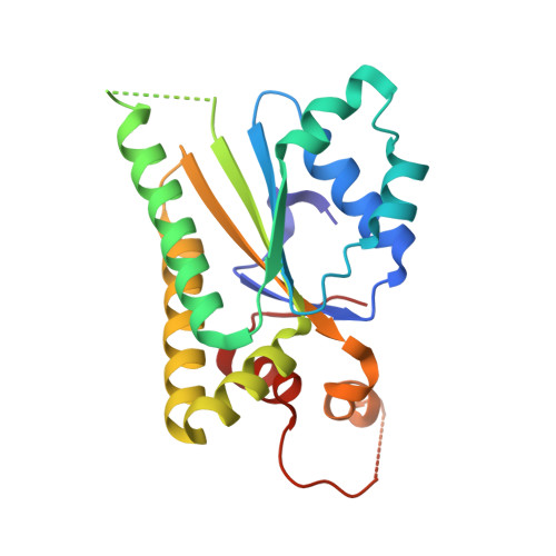
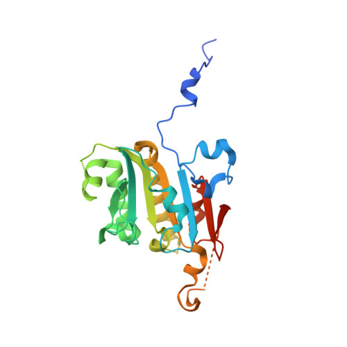
    The yeast Shu complex, consisting of the proteins Shu1, Shu2, Psy3, and Csm2, maintains genomic stability by coupling post-replication repair to homologous recombination. However, a lack of biochemical and structural information on the Shu proteins precludes revealing their precise roles within the pathway. Here, we report on the 1.9-Å crystal structure of the Psy3-Csm2 complex. The crystal structure shows that Psy3 forms a heterodimer with Csm2 mainly through a hydrophobic core. Unexpectedly, Psy3 and Csm2 share a similar architecture that closely resembles the ATPase core domain of Rad51. The L2 loop present in Psy3 and Csm2 is similar to that of Rad51 and confers the DNA binding activity of the Shu complex. As with Rad51, the Shu complex appears to form a nucleoprotein filament by binding nonspecifically to DNA. Structure-based mutagenesis studies have demonstrated that the DNA binding activity of the Shu complex is essential for repair of the methyl methanesulfonate-induced DNA damage. Our findings provide good foundations for the understanding of the Srs2 regulation by the Shu complex.


  • Organizational Affiliation

    Hefei National Laboratory for Physical Sciences at Microscale and School of Life Sciences, University of Science and Technology of China, Hefei, Anhui 230026, China.


Macromolecules
Find similar proteins by:  (by identity cutoff)  |  3D Structure
Entity ID: 1
MoleculeChains Sequence LengthOrganismDetailsImage
Chromosome segregation in meiosis protein 2213Saccharomyces cerevisiae S288CMutation(s): 0 
Gene Names: CSM2
UniProt
Find proteins for P40465 (Saccharomyces cerevisiae (strain ATCC 204508 / S288c))
Explore P40465 
Go to UniProtKB:  P40465
Entity Groups  
Sequence Clusters30% Identity50% Identity70% Identity90% Identity95% Identity100% Identity
UniProt GroupP40465
Sequence Annotations
Expand
  • Reference Sequence
Find similar proteins by:  (by identity cutoff)  |  3D Structure
Entity ID: 2
MoleculeChains Sequence LengthOrganismDetailsImage
Platinum sensitivity protein 3245Saccharomyces cerevisiae S288CMutation(s): 0 
Gene Names: PSY3
UniProt
Find proteins for Q12318 (Saccharomyces cerevisiae (strain ATCC 204508 / S288c))
Explore Q12318 
Go to UniProtKB:  Q12318
Entity Groups  
Sequence Clusters30% Identity50% Identity70% Identity90% Identity95% Identity100% Identity
UniProt GroupQ12318
Sequence Annotations
Expand
  • Reference Sequence
Experimental Data & Validation

Experimental Data

  • Method: X-RAY DIFFRACTION
  • Resolution: 1.90 Å
  • R-Value Free: 0.230 
  • R-Value Work: 0.192 
  • R-Value Observed: 0.194 
  • Space Group: C 1 2 1
Unit Cell:
Length ( Å )Angle ( ˚ )
a = 135.123α = 90
b = 50.433β = 103.17
c = 76.833γ = 90
Software Package:
Software NamePurpose
HKL-2000data collection
SOLVEphasing
PHENIXrefinement
HKL-2000data reduction
SCALEPACKdata scaling

Structure Validation

View Full Validation Report


Entry History 

Deposition Data

Revision History  (Full details and data files)

  • Version 1.0: 2012-04-11
    Type: Initial release
  • Version 1.1: 2013-06-26
    Changes: Database references
  • Version 1.2: 2024-03-20
    Changes: Data collection, Database references, Derived calculations