4DQO

Crystal Structure of PG16 Fab in Complex with V1V2 Region from HIV-1 strain ZM109


Experimental Data Snapshot

  • Method: X-RAY DIFFRACTION
  • Resolution: 2.44 Å
  • R-Value Free: 0.230 
  • R-Value Work: 0.201 
  • R-Value Observed: 0.203 

wwPDB Validation   3D Report Full Report


This is version 3.3 of the entry. See complete history


Literature

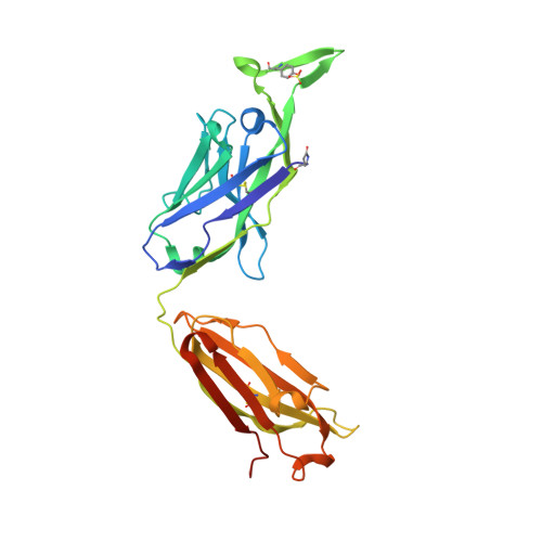
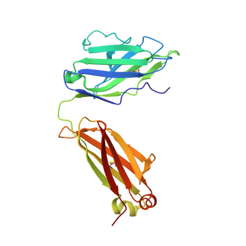
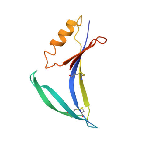
Structural basis for diverse N-glycan recognition by HIV-1-neutralizing V1-V2-directed antibody PG16.

Pancera, M.Shahzad-Ul-Hussan, S.Doria-Rose, N.A.McLellan, J.S.Bailer, R.T.Dai, K.Loesgen, S.Louder, M.K.Staupe, R.P.Yang, Y.Zhang, B.Parks, R.Eudailey, J.Lloyd, K.E.Blinn, J.Alam, S.M.Haynes, B.F.Amin, M.N.Wang, L.X.Burton, D.R.Koff, W.C.Nabel, G.J.Mascola, J.R.Bewley, C.A.Kwong, P.D.

(2013) Nat Struct Mol Biol 20: 804-813

  • DOI: https://doi.org/10.1038/nsmb.2600
  • Primary Citation of Related Structures:  
    4DQO

  • PubMed Abstract: 

    HIV-1 uses a diverse N-linked-glycan shield to evade recognition by antibody. Select human antibodies, such as the clonally related PG9 and PG16, recognize glycopeptide epitopes in the HIV-1 V1-V2 region and penetrate this shield, but their ability to accommodate diverse glycans is unclear. Here we report the structure of antibody PG16 bound to a scaffolded V1-V2, showing an epitope comprising both high mannose-type and complex-type N-linked glycans. We combined structure, NMR and mutagenesis analyses to characterize glycan recognition by PG9 and PG16. Three PG16-specific residues, arginine, serine and histidine (RSH), were critical for binding sialic acid on complex-type glycans, and introduction of these residues into PG9 produced a chimeric antibody with enhanced HIV-1 neutralization. Although HIV-1-glycan diversity facilitates evasion, antibody somatic diversity can overcome this and can provide clues to guide the design of modified antibodies with enhanced neutralization.


  • Organizational Affiliation

    Vaccine Research Center, National Institute of Allergy and Infectious Diseases, US National Institutes of Health, Bethesda, Maryland, USA.


Macromolecules
Find similar proteins by:  (by identity cutoff)  |  3D Structure
Entity ID: 1
MoleculeChains Sequence LengthOrganismDetailsImage
PG16 Fab Heavy ChainA [auth H]246Homo sapiensMutation(s): 0 
Entity Groups  
Sequence Clusters30% Identity50% Identity70% Identity90% Identity95% Identity100% Identity
Sequence Annotations
Expand
  • Reference Sequence
Find similar proteins by:  (by identity cutoff)  |  3D Structure
Entity ID: 2
MoleculeChains Sequence LengthOrganismDetailsImage
PG16 Fab Light ChainB [auth L]216Homo sapiensMutation(s): 0 
Entity Groups  
Sequence Clusters30% Identity50% Identity70% Identity90% Identity95% Identity100% Identity
Sequence Annotations
Expand
  • Reference Sequence
Find similar proteins by:  (by identity cutoff)  |  3D Structure
Entity ID: 3
MoleculeChains Sequence LengthOrganismDetailsImage
1FD6-V1V2 scaffold ZM109 HIV-1 strain124Human immunodeficiency virus 1Mutation(s): 0 
UniProt
Find proteins for Q6TCP8 (Human immunodeficiency virus 1)
Explore Q6TCP8 
Go to UniProtKB:  Q6TCP8
Entity Groups  
Sequence Clusters30% Identity50% Identity70% Identity90% Identity95% Identity100% Identity
UniProt GroupQ6TCP8
Sequence Annotations
Expand
  • Reference Sequence
Oligosaccharides

Help

Entity ID: 4
MoleculeChains Length2D Diagram Glycosylation3D Interactions
alpha-D-mannopyranose-(1-3)-[alpha-D-mannopyranose-(1-6)]alpha-D-mannopyranose-(1-6)-[alpha-D-mannopyranose-(1-3)]beta-D-mannopyranose-(1-4)-2-acetamido-2-deoxy-beta-D-glucopyranose-(1-4)-2-acetamido-2-deoxy-beta-D-glucopyranoseD [auth A]7N-Glycosylation
Glycosylation Resources
GlyTouCan:  G55220VL
GlyCosmos:  G55220VL
GlyGen:  G55220VL
Entity ID: 5
MoleculeChains Length2D Diagram Glycosylation3D Interactions
N-acetyl-alpha-neuraminic acid-(2-6)-beta-D-galactopyranose-(1-4)-2-acetamido-2-deoxy-beta-D-glucopyranose-(1-2)-alpha-D-mannopyranose-(1-3)-[alpha-D-mannopyranose-(1-3)-[alpha-D-mannopyranose-(1-6)]alpha-D-mannopyranose-(1-6)]beta-D-mannopyranose-(1-4)-2-acetamido-2-deoxy-beta-D-glucopyranose-(1-4)-2-acetamido-2-deoxy-beta-D-glucopyranoseE [auth B]10N-Glycosylation
Glycosylation Resources
GlyTouCan:  G04576KS
GlyCosmos:  G04576KS
GlyGen:  G04576KS
Small Molecules
Modified Residues  2 Unique
IDChains TypeFormula2D DiagramParent
PCA
Query on PCA
A [auth H]L-PEPTIDE LINKINGC5 H7 N O3GLN
TYS
Query on TYS
A [auth H]L-PEPTIDE LINKINGC9 H11 N O6 STYR
Experimental Data & Validation

Experimental Data

  • Method: X-RAY DIFFRACTION
  • Resolution: 2.44 Å
  • R-Value Free: 0.230 
  • R-Value Work: 0.201 
  • R-Value Observed: 0.203 
  • Space Group: C 2 2 21
Unit Cell:
Length ( Å )Angle ( ˚ )
a = 81.054α = 90
b = 207.561β = 90
c = 87.617γ = 90
Software Package:
Software NamePurpose
HKL-2000data collection
PHASERphasing
PHENIXrefinement
HKL-2000data reduction
HKL-2000data scaling

Structure Validation

View Full Validation Report


Entry History 

Deposition Data

Revision History  (Full details and data files)

  • Version 1.0: 2013-03-06
    Type: Initial release
  • Version 1.1: 2013-05-08
    Changes: Derived calculations, Other
  • Version 1.2: 2013-06-05
    Changes: Database references
  • Version 1.3: 2013-07-24
    Changes: Database references
  • Version 2.0: 2019-12-25
    Changes: Data collection, Derived calculations, Polymer sequence
  • Version 3.0: 2020-07-29
    Type: Remediation
    Reason: Carbohydrate remediation
    Changes: Advisory, Atomic model, Data collection, Derived calculations, Structure summary
  • Version 3.1: 2021-05-19
    Changes: Derived calculations, Source and taxonomy, Structure summary
  • Version 3.2: 2023-09-13
    Changes: Data collection, Database references, Refinement description
  • Version 3.3: 2023-12-06
    Changes: Data collection