4DMZ

PelD 156-455 from Pseudomonas aeruginosa PA14, apo form


Experimental Data Snapshot

  • Method: X-RAY DIFFRACTION
  • Resolution: 2.10 Å
  • R-Value Free: 0.238 
  • R-Value Work: 0.196 
  • R-Value Observed: 0.198 

wwPDB Validation   3D Report Full Report


This is version 1.3 of the entry. See complete history


Literature

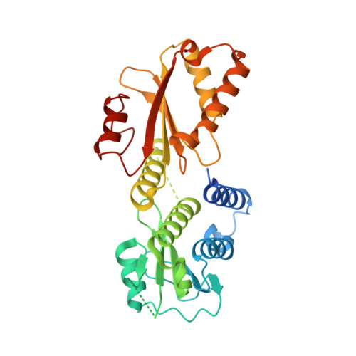
Structure of the Cytoplasmic Region of PelD, a Degenerate Diguanylate Cyclase Receptor That Regulates Exopolysaccharide Production in Pseudomonas aeruginosa.

Whitney, J.C.Colvin, K.M.Marmont, L.S.Robinson, H.Parsek, M.R.Howell, P.L.

(2012) J Biol Chem 287: 23582-23593

  • DOI: https://doi.org/10.1074/jbc.M112.375378
  • Primary Citation of Related Structures:  
    4DMZ, 4DN0

  • PubMed Abstract: 

    High cellular concentrations of bis-(3',5')-cyclic dimeric guanosine mono-phosphate (c-di-GMP) regulate a diverse range of phenotypes in bacteria including biofilm development. The opportunistic pathogen Pseudomonas aeruginosa produces the PEL polysaccharide to form a biofilm at the air-liquid interface of standing cultures. Among the proteins required for PEL polysaccharide production, PelD has been identified as a membrane-bound c-di-GMP-specific receptor. In this work, we present the x-ray crystal structure of a soluble cytoplasmic region of PelD in its apo and c-di-GMP complexed forms. The structure of PelD reveals an N-terminal GAF domain and a C-terminal degenerate GGDEF domain, the latter of which binds dimeric c-di-GMP at an RXXD motif that normally serves as an allosteric inhibition site for active diguanylate cyclases. Using isothermal titration calorimetry, we demonstrate that PelD binds c-di-GMP with low micromolar affinity and that mutation of residues involved in binding not only decreases the affinity of this interaction but also abrogates PEL-specific phenotypes in vivo. Bioinformatics analysis of the juxtamembrane region of PelD suggests that it contains an α-helical stalk region that connects the soluble region to the transmembrane domains and that similarly to other GAF domain containing proteins, this region likely forms a coiled-coil motif that mediates dimerization. PelD with Alg44 and BcsA of the alginate and cellulose secretion systems, respectively, collectively constitute a group of c-di-GMP receptors that appear to regulate exopolysaccharide assembly at the protein level through activation of their associated glycosyl transferases.


  • Organizational Affiliation

    Program in Molecular Structure and Function, Hospital for Sick Children, Toronto, Ontario M5G 1X8, Canada.


Macromolecules
Find similar proteins by:  (by identity cutoff)  |  3D Structure
Entity ID: 1
MoleculeChains Sequence LengthOrganismDetailsImage
Putative uncharacterized protein pelD
A, B
321Pseudomonas aeruginosa UCBPP-PA14Mutation(s): 0 
Gene Names: pelDPA14_24510
UniProt
Find proteins for A0A0H2ZD98 (Pseudomonas aeruginosa (strain UCBPP-PA14))
Explore A0A0H2ZD98 
Go to UniProtKB:  A0A0H2ZD98
Entity Groups  
Sequence Clusters30% Identity50% Identity70% Identity90% Identity95% Identity100% Identity
UniProt GroupA0A0H2ZD98
Sequence Annotations
Expand
  • Reference Sequence
Small Molecules
Ligands 3 Unique
IDChains Name / Formula / InChI Key2D Diagram3D Interactions
TRS
Query on TRS

Download Ideal Coordinates CCD File 
Z [auth B]2-AMINO-2-HYDROXYMETHYL-PROPANE-1,3-DIOL
C4 H12 N O3
LENZDBCJOHFCAS-UHFFFAOYSA-O
EDO
Query on EDO

Download Ideal Coordinates CCD File 
Q [auth A],
R [auth A],
Y [auth B]
1,2-ETHANEDIOL
C2 H6 O2
LYCAIKOWRPUZTN-UHFFFAOYSA-N
MG
Query on MG

Download Ideal Coordinates CCD File 
C [auth A]
D [auth A]
E [auth A]
F [auth A]
G [auth A]
C [auth A],
D [auth A],
E [auth A],
F [auth A],
G [auth A],
H [auth A],
I [auth A],
J [auth A],
K [auth A],
L [auth A],
M [auth A],
N [auth A],
O [auth A],
P [auth A],
S [auth B],
T [auth B],
U [auth B],
V [auth B],
W [auth B],
X [auth B]
MAGNESIUM ION
Mg
JLVVSXFLKOJNIY-UHFFFAOYSA-N
Experimental Data & Validation

Experimental Data

  • Method: X-RAY DIFFRACTION
  • Resolution: 2.10 Å
  • R-Value Free: 0.238 
  • R-Value Work: 0.196 
  • R-Value Observed: 0.198 
  • Space Group: P 21 21 2
Unit Cell:
Length ( Å )Angle ( ˚ )
a = 88.511α = 90
b = 113.346β = 90
c = 61.846γ = 90
Software Package:
Software NamePurpose
HKL-2000data collection
SOLVEphasing
PHENIXrefinement
HKL-2000data reduction
HKL-2000data scaling

Structure Validation

View Full Validation Report


Entry History 

Deposition Data

Revision History  (Full details and data files)

  • Version 1.0: 2012-05-23
    Type: Initial release
  • Version 1.1: 2012-05-30
    Changes: Database references
  • Version 1.2: 2012-07-25
    Changes: Database references
  • Version 1.3: 2024-02-28
    Changes: Data collection, Database references, Derived calculations