4DCQ

Crystal Structure of the Fab Fragment of 3B5H10, an Antibody-Specific for Extended Polyglutamine Repeats (orthorhombic form)


Experimental Data Snapshot

  • Method: X-RAY DIFFRACTION
  • Resolution: 1.94 Å
  • R-Value Free: 0.246 
  • R-Value Work: 0.185 
  • R-Value Observed: 0.188 

wwPDB Validation   3D Report Full Report


This is version 1.2 of the entry. See complete history


Literature

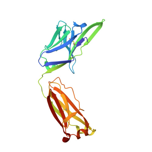
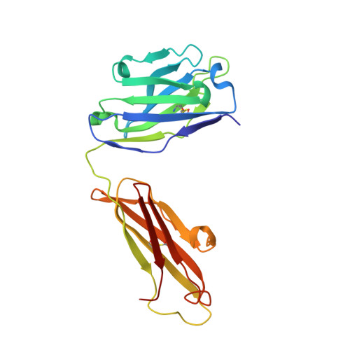
Disease-associated polyglutamine stretches in monomeric huntingtin adopt a compact structure.

Peters-Libeu, C.Miller, J.Rutenber, E.Newhouse, Y.Krishnan, P.Cheung, K.Hatters, D.Brooks, E.Widjaja, K.Tran, T.Mitra, S.Arrasate, M.Mosquera, L.A.Taylor, D.Weisgraber, K.H.Finkbeiner, S.

(2012) J Mol Biol 421: 587-600

  • DOI: https://doi.org/10.1016/j.jmb.2012.01.034
  • Primary Citation of Related Structures:  
    3S96, 4DCQ

  • PubMed Abstract: 

    Abnormal polyglutamine (polyQ) tracts are the only common feature in nine proteins that each cause a dominant neurodegenerative disorder. In Huntington's disease, tracts longer than 36 glutamines in the protein huntingtin (htt) cause degeneration. In situ, monoclonal antibody 3B5H10 binds to different htt fragments in neurons in proportion to their toxicity. Here, we determined the structure of 3B5H10 Fab to 1.9 Å resolution by X-ray crystallography. Modeling demonstrates that the paratope forms a groove suitable for binding two β-rich polyQ strands. Using small-angle X-ray scattering, we confirmed that the polyQ epitope recognized by 3B5H10 is a compact two-stranded hairpin within monomeric htt and is abundant in htt fragments unbound to antibody. Thus, disease-associated polyQ stretches preferentially adopt compact conformations. Since 3B5H10 binding predicts degeneration, this compact polyQ structure may be neurotoxic.


  • Organizational Affiliation

    Gladstone Institute of Neurological Disease, San Francisco, CA 94158, USA.


Macromolecules
Find similar proteins by:  (by identity cutoff)  |  3D Structure
Entity ID: 1
MoleculeChains Sequence LengthOrganismDetailsImage
3B5H10 FAB Light Chain218Mus musculusMutation(s): 0 
Entity Groups  
Sequence Clusters30% Identity50% Identity70% Identity90% Identity95% Identity100% Identity
Sequence Annotations
Expand
  • Reference Sequence
Find similar proteins by:  (by identity cutoff)  |  3D Structure
Entity ID: 2
MoleculeChains Sequence LengthOrganismDetailsImage
3B5H10 FAB Heavy Chain218Mus musculusMutation(s): 0 
Entity Groups  
Sequence Clusters30% Identity50% Identity70% Identity90% Identity95% Identity100% Identity
Sequence Annotations
Expand
  • Reference Sequence
Experimental Data & Validation

Experimental Data

  • Method: X-RAY DIFFRACTION
  • Resolution: 1.94 Å
  • R-Value Free: 0.246 
  • R-Value Work: 0.185 
  • R-Value Observed: 0.188 
  • Space Group: P 21 21 2
Unit Cell:
Length ( Å )Angle ( ˚ )
a = 78.068α = 90
b = 121.815β = 90
c = 42.012γ = 90
Software Package:
Software NamePurpose
MOSFLMdata reduction
SCALEPACKdata scaling
PHASERphasing
REFMACrefinement
PDB_EXTRACTdata extraction
ADSCdata collection

Structure Validation

View Full Validation Report


Entry History 

Deposition Data

Revision History  (Full details and data files)

  • Version 1.0: 2012-02-22
    Type: Initial release
  • Version 1.1: 2012-08-08
    Changes: Database references
  • Version 1.2: 2023-09-13
    Changes: Data collection, Database references, Derived calculations, Refinement description