4D73

X-ray structure of a peroxiredoxin


Experimental Data Snapshot

  • Method: X-RAY DIFFRACTION
  • Resolution: 1.80 Å
  • R-Value Free: 0.195 
  • R-Value Work: 0.152 
  • R-Value Observed: 0.154 

wwPDB Validation   3D Report Full Report


This is version 1.5 of the entry. See complete history


Literature

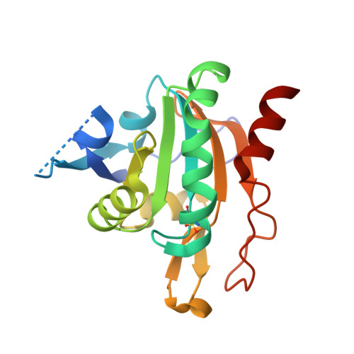
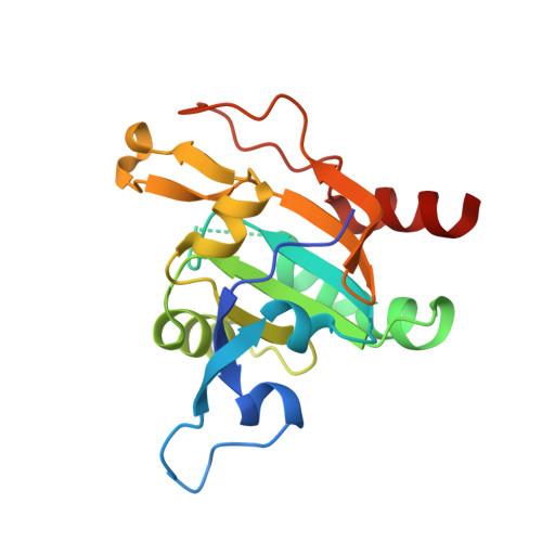
Plasmodium Falciparum Antioxidant Protein Reveals a Novel Mechanism for Balancing Turnover and Inactivation of Peroxiredoxins

Staudacher, V.Djuika, C.F.Koduka, J.Schlossarek, S.Kopp, J.Buechler, M.Lanzer, M.Deponte, M.

(2015) Free Radic Biol Med 85: 228

  • DOI: https://doi.org/10.1016/j.freeradbiomed.2015.04.030
  • Primary Citation of Related Structures:  
    4D73

  • PubMed Abstract: 

    Life under aerobic conditions has shaped peroxiredoxins (Prx) as ubiquitous thiol-dependent hydroperoxidases and redox sensors. Structural features that balance the catalytically active or inactive redox states of Prx, and, therefore, their hydroperoxidase or sensor function, have so far been analyzed predominantly for Prx1-type enzymes. Here we identify and characterize two modulatory residues of the Prx5-type model enzyme PfAOP from the malaria parasite Plasmodium falciparum. Gain- and loss-of-function mutants reveal a correlation between the enzyme parameters and the inactivation susceptibility of PfAOP with the size of residue 109 and the presence or absence of a catalytically relevant but nonessential cysteine residue. Based on our kinetic data and the crystal structure of PfAOP(L109M), we suggest a novel mechanism for balancing the hydroperoxidase activity and inactivation susceptibility of Prx5-type enzymes. Our study provides unexpected insights into Prx structure-function relationships and contributes to our understanding of what makes Prx good enzymes or redox sensors.


  • Organizational Affiliation

    Department of Parasitology, Ruprecht-Karls University, D-69120 Heidelberg, Germany.


Macromolecules
Find similar proteins by:  (by identity cutoff)  |  3D Structure
Entity ID: 1
MoleculeChains Sequence LengthOrganismDetailsImage
1-CYS PEROXIREDOXIN180Plasmodium falciparumMutation(s): 1 
EC: 1.11.1.15
UniProt
Find proteins for Q5MYR6 (Plasmodium falciparum (isolate 3D7))
Explore Q5MYR6 
Go to UniProtKB:  Q5MYR6
Entity Groups  
Sequence Clusters30% Identity50% Identity70% Identity90% Identity95% Identity100% Identity
UniProt GroupQ5MYR6
Sequence Annotations
Expand
  • Reference Sequence
Find similar proteins by:  (by identity cutoff)  |  3D Structure
Entity ID: 2
MoleculeChains Sequence LengthOrganismDetailsImage
1-CYS PEROXIREDOXIN180Plasmodium falciparumMutation(s): 1 
EC: 1.11.1.15
UniProt
Find proteins for Q5MYR6 (Plasmodium falciparum (isolate 3D7))
Explore Q5MYR6 
Go to UniProtKB:  Q5MYR6
Entity Groups  
Sequence Clusters30% Identity50% Identity70% Identity90% Identity95% Identity100% Identity
UniProt GroupQ5MYR6
Sequence Annotations
Expand
  • Reference Sequence
Small Molecules
Modified Residues  1 Unique
IDChains TypeFormula2D DiagramParent
OCS
Query on OCS
A
L-PEPTIDE LINKINGC3 H7 N O5 SCYS
Experimental Data & Validation

Experimental Data

  • Method: X-RAY DIFFRACTION
  • Resolution: 1.80 Å
  • R-Value Free: 0.195 
  • R-Value Work: 0.152 
  • R-Value Observed: 0.154 
  • Space Group: P 21 21 21
Unit Cell:
Length ( Å )Angle ( ˚ )
a = 41.419α = 90
b = 77.366β = 90
c = 109.751γ = 90
Software Package:
Software NamePurpose
PHENIXrefinement
XDSdata reduction
Aimlessdata scaling
PHASERphasing

Structure Validation

View Full Validation Report


Entry History 

Deposition Data

Revision History  (Full details and data files)

  • Version 1.0: 2015-05-13
    Type: Initial release
  • Version 1.1: 2015-05-20
    Changes: Database references
  • Version 1.2: 2015-08-05
    Changes: Database references
  • Version 1.3: 2019-03-06
    Changes: Data collection, Derived calculations, Experimental preparation, Other
  • Version 1.4: 2019-05-08
    Changes: Data collection, Experimental preparation
  • Version 1.5: 2023-12-20
    Changes: Data collection, Database references, Other, Refinement description