4D02

The crystallographic structure of Flavorubredoxin from Escherichia coli


Experimental Data Snapshot

  • Method: X-RAY DIFFRACTION
  • Resolution: 1.75 Å
  • R-Value Work: 0.141 
  • R-Value Observed: 0.141 

wwPDB Validation   3D Report Full Report


Ligand Structure Quality Assessment 


This is version 1.3 of the entry. See complete history


Literature

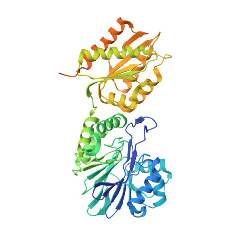
Structure of Escherichia Coli Flavodiiron Nitric Oxide Reductase.

Romao, C.V.Vicente, J.B.Borges, P.T.Victor, B.L.Lamosa, P.Silva, E.Bandeiras, T.M.Soares, C.M.Carrondo, M.A.Turner, D.Teixeira, M.Frazao, C.

(2016) J Mol Biol 428: 4686

  • DOI: https://doi.org/10.1016/j.jmb.2016.10.008
  • Primary Citation of Related Structures:  
    4D02, 5LLD, 5LMC

  • PubMed Abstract: 

    Flavodiiron proteins (FDPs) are present in organisms from all domains of life and have been described so far to be involved in the detoxification of oxygen or nitric oxide (NO), acting as O 2 and/or NO reductases. The Escherichia coli FDP, named flavorubredoxin (FlRd), is the most extensively studied FDP. Biochemical and in vivo studies revealed that FlRd is involved in NO detoxification as part of the bacterial defense mechanisms against reactive nitrogen species. E. coli FlRd has a clear preference for NO as a substrate in vitro, exhibiting a very low reactivity toward O 2 . To contribute to the understanding of the structural features defining this substrate selectivity, we determined the crystallographic structure of E. coli FlRd, both in the isolated and reduced states. The overall tetrameric structure revealed a highly conserved flavodiiron core domain, with a metallo-β-lactamase-like domain containing a diiron center, and a flavodoxin domain with a flavin mononucleotide cofactor. The metal center in the oxidized state has a μ-hydroxo bridge coordinating the two irons, while in the reduced state, this moiety is not detected. Since only the flavodiiron domain was observed in these crystal structures, the structure of the rubredoxin domain was determined by NMR. Tunnels for the substrates were identified, and through molecular dynamics simulations, no differences for O 2 or NO permeation were found. The present data represent the first structure for a NO-selective FDP.


  • Organizational Affiliation

    Instituto de Tecnologia Química e Biológica António Xavier, ITQB NOVA, Av. da República, 2780-157 Oeiras, Portugal.


Macromolecules
Find similar proteins by:  (by identity cutoff)  |  3D Structure
Entity ID: 1
MoleculeChains Sequence LengthOrganismDetailsImage
ANAEROBIC NITRIC OXIDE REDUCTASE FLAVORUBREDOXIN479Escherichia coli K-12Mutation(s): 0 
UniProt
Find proteins for Q46877 (Escherichia coli (strain K12))
Explore Q46877 
Go to UniProtKB:  Q46877
Entity Groups  
Sequence Clusters30% Identity50% Identity70% Identity90% Identity95% Identity100% Identity
UniProt GroupQ46877
Sequence Annotations
Expand
  • Reference Sequence
Small Molecules
Ligands 6 Unique
IDChains Name / Formula / InChI Key2D Diagram3D Interactions
FMN
Query on FMN

Download Ideal Coordinates CCD File 
B [auth A]FLAVIN MONONUCLEOTIDE
C17 H21 N4 O9 P
FVTCRASFADXXNN-SCRDCRAPSA-N
PO4
Query on PO4

Download Ideal Coordinates CCD File 
F [auth A],
G [auth A]
PHOSPHATE ION
O4 P
NBIIXXVUZAFLBC-UHFFFAOYSA-K
GOL
Query on GOL

Download Ideal Coordinates CCD File 
J [auth A],
K [auth A]
GLYCEROL
C3 H8 O3
PEDCQBHIVMGVHV-UHFFFAOYSA-N
FE
Query on FE

Download Ideal Coordinates CCD File 
C [auth A],
D [auth A]
FE (III) ION
Fe
VTLYFUHAOXGGBS-UHFFFAOYSA-N
CL
Query on CL

Download Ideal Coordinates CCD File 
H [auth A],
I [auth A]
CHLORIDE ION
Cl
VEXZGXHMUGYJMC-UHFFFAOYSA-M
O
Query on O

Download Ideal Coordinates CCD File 
E [auth A]OXYGEN ATOM
O
XLYOFNOQVPJJNP-UHFFFAOYSA-N
Experimental Data & Validation

Experimental Data

  • Method: X-RAY DIFFRACTION
  • Resolution: 1.75 Å
  • R-Value Work: 0.141 
  • R-Value Observed: 0.141 
  • Space Group: P 6 2 2
Unit Cell:
Length ( Å )Angle ( ˚ )
a = 149.529α = 90
b = 149.529β = 90
c = 94.498γ = 120
Software Package:
Software NamePurpose
PHENIXrefinement
XDSdata reduction
XDSdata scaling
SHELXCDphasing
SHELXEmodel building

Structure Validation

View Full Validation Report


Ligand Structure Quality Assessment 


Entry History 

Deposition Data

Revision History  (Full details and data files)

  • Version 1.0: 2014-09-03
    Type: Initial release
  • Version 1.1: 2014-10-22
    Changes: Database references
  • Version 1.2: 2017-02-08
    Changes: Database references
  • Version 1.3: 2019-05-08
    Changes: Data collection, Experimental preparation, Other