4C2K

Crystal structure of human mitochondrial 3-ketoacyl-CoA thiolase


Experimental Data Snapshot

  • Method: X-RAY DIFFRACTION
  • Resolution: 2.00 Å
  • R-Value Free: 0.229 
  • R-Value Work: 0.195 
  • R-Value Observed: 0.197 

wwPDB Validation   3D Report Full Report


This is version 1.3 of the entry. See complete history


Literature

The Crystal Structure of Human Mitochondrial 3-Ketoacyl-Coa Thiolase (T1): Insight Into the Reaction Mechanism of its Thiolase and Thioesterase Activities

Kiema, T.-R.Harijan, R.K.Strozyk, M.Fukao, T.Alexson, S.E.H.Wierenga, R.K.

(2014) Acta Crystallogr D Biol Crystallogr 70: 3212

  • DOI: https://doi.org/10.1107/S1399004714023827
  • Primary Citation of Related Structures:  
    4C2J, 4C2K

  • PubMed Abstract: 

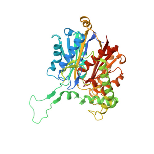
    Crystal structures of human mitochondrial 3-ketoacyl-CoA thiolase (hT1) in the apo form and in complex with CoA have been determined at 2.0 Å resolution. The structures confirm the tetrameric quaternary structure of this degradative thiolase. The active site is surprisingly similar to the active site of the Zoogloea ramigera biosynthetic tetrameric thiolase (PDB entries 1dm3 and 1m1o) and different from the active site of the peroxisomal dimeric degradative thiolase (PDB entries 1afw and 2iik). A cavity analysis suggests a mode of binding for the fatty-acyl tail in a tunnel lined by the Nβ2-Nα2 loop of the adjacent subunit and the Lα1 helix of the loop domain. Soaking of the apo hT1 crystals with octanoyl-CoA resulted in a crystal structure in complex with CoA owing to the intrinsic acyl-CoA thioesterase activity of hT1. Solution studies confirm that hT1 has low acyl-CoA thioesterase activity for fatty acyl-CoA substrates. The fastest rate is observed for the hydrolysis of butyryl-CoA. It is also shown that T1 has significant biosynthetic thiolase activity, which is predicted to be of physiological importance.


  • Organizational Affiliation

    Faculty of Biochemistry and Molecular Medicine, Biocenter Oulu, University of Oulu, PO Box 3000, FIN-90014 Oulu, Finland.


Macromolecules
Find similar proteins by:  (by identity cutoff)  |  3D Structure
Entity ID: 1
MoleculeChains Sequence LengthOrganismDetailsImage
3-KETOACYL-COA THIOLASE, MITOCHONDRIAL
A, B, C, D
417Homo sapiensMutation(s): 0 
EC: 2.3.1.16
UniProt & NIH Common Fund Data Resources
Find proteins for P42765 (Homo sapiens)
Explore P42765 
Go to UniProtKB:  P42765
PHAROS:  P42765
GTEx:  ENSG00000167315 
Entity Groups  
Sequence Clusters30% Identity50% Identity70% Identity90% Identity95% Identity100% Identity
UniProt GroupP42765
Sequence Annotations
Expand
  • Reference Sequence
Small Molecules
Ligands 3 Unique
IDChains Name / Formula / InChI Key2D Diagram3D Interactions
MES
Query on MES

Download Ideal Coordinates CCD File 
DA [auth D],
I [auth A],
O [auth B],
P [auth B]
2-(N-MORPHOLINO)-ETHANESULFONIC ACID
C6 H13 N O4 S
SXGZJKUKBWWHRA-UHFFFAOYSA-N
DTT
Query on DTT

Download Ideal Coordinates CCD File 
H [auth A]2,3-DIHYDROXY-1,4-DITHIOBUTANE
C4 H10 O2 S2
VHJLVAABSRFDPM-IMJSIDKUSA-N
EDO
Query on EDO

Download Ideal Coordinates CCD File 
AA [auth D]
BA [auth D]
CA [auth D]
E [auth A]
F [auth A]
AA [auth D],
BA [auth D],
CA [auth D],
E [auth A],
F [auth A],
G [auth A],
J [auth B],
K [auth B],
L [auth B],
M [auth B],
N [auth B],
Q [auth C],
R [auth C],
S [auth C],
T [auth C],
U [auth C],
V [auth C],
W [auth C],
X [auth D],
Y [auth D],
Z [auth D]
1,2-ETHANEDIOL
C2 H6 O2
LYCAIKOWRPUZTN-UHFFFAOYSA-N
Experimental Data & Validation

Experimental Data

  • Method: X-RAY DIFFRACTION
  • Resolution: 2.00 Å
  • R-Value Free: 0.229 
  • R-Value Work: 0.195 
  • R-Value Observed: 0.197 
  • Space Group: P 1 21 1
Unit Cell:
Length ( Å )Angle ( ˚ )
a = 54.73α = 90
b = 196.77β = 103.78
c = 79.65γ = 90
Software Package:
Software NamePurpose
REFMACrefinement
XDSdata reduction
PHASERphasing

Structure Validation

View Full Validation Report


Entry History 

Deposition Data

Revision History  (Full details and data files)

  • Version 1.0: 2014-09-03
    Type: Initial release
  • Version 1.1: 2014-12-10
    Changes: Database references
  • Version 1.2: 2015-07-22
    Changes: Database references
  • Version 1.3: 2023-12-20
    Changes: Data collection, Database references, Derived calculations, Other, Refinement description