4B2N

Latex Oxygenase RoxA


Experimental Data Snapshot

  • Method: X-RAY DIFFRACTION
  • Resolution: 1.80 Å
  • R-Value Free: 0.220 
  • R-Value Work: 0.168 
  • R-Value Observed: 0.171 

wwPDB Validation   3D Report Full Report


Ligand Structure Quality Assessment 


This is version 2.2 of the entry. See complete history


Literature

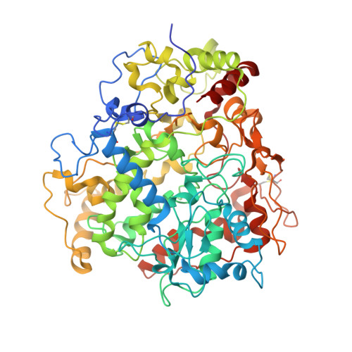
Structure of the processive rubber oxygenase RoxA from Xanthomonas sp.

Seidel, J.Schmitt, G.Hoffmann, M.Jendrossek, D.Einsle, O.

(2013) Proc Natl Acad Sci U S A 110: 13833-13838

  • DOI: https://doi.org/10.1073/pnas.1305560110
  • Primary Citation of Related Structures:  
    4B2N

  • PubMed Abstract: 

    Rubber oxygenase A (RoxA) is one of only two known enzymes able to catalyze the oxidative cleavage of latex for biodegradation. RoxA acts as a processive dioxygenase to yield the predominant product 12-oxo-4,8-dimethyl-trideca-4,8-diene-1-al (ODTD), a tri-isoprene unit. Here we present a structural analysis of RoxA from Xanthomonas sp. strain 35Y at a resolution of 1.8 Å. The enzyme is a 75-kDa diheme c-type cytochrome with an unusually low degree of secondary structure. Analysis of the heme group arrangement and peptide chain topology of RoxA confirmed a distant kinship with diheme peroxidases of the CcpA family, but the proteins are functionally distinct, and the extracellular RoxA has evolved to have twice the molecular mass by successively accumulating extensions of peripheral loops. RoxA incorporates both oxygen atoms of its cosubstrate dioxygen into the rubber cleavage product ODTD, and we show that RoxA is isolated with O2 stably bound to the active site heme iron. Activation and cleavage of O2 require binding of polyisoprene, and thus the substrate needs to use hydrophobic access channels to reach the deeply buried active site of RoxA. The location and nature of these channels support a processive mechanism of latex cleavage.


  • Organizational Affiliation

    Lehrstuhl für Biochemie, Institut für Biochemie, Albert-Ludwigs-Universität Freiburg, 79104 Freiburg, Germany.


Macromolecules
Find similar proteins by:  (by identity cutoff)  |  3D Structure
Entity ID: 1
MoleculeChains Sequence LengthOrganismDetailsImage
70 KDA PROTEIN
A, B
662Xanthomonas sp. 35YMutation(s): 0 
UniProt
Find proteins for Q7X0P3 (Xanthomonas sp. 35Y)
Explore Q7X0P3 
Go to UniProtKB:  Q7X0P3
Entity Groups  
Sequence Clusters30% Identity50% Identity70% Identity90% Identity95% Identity100% Identity
UniProt GroupQ7X0P3
Sequence Annotations
Expand
  • Reference Sequence
Small Molecules
Ligands 3 Unique
IDChains Name / Formula / InChI Key2D Diagram3D Interactions
HEC
Query on HEC

Download Ideal Coordinates CCD File 
C [auth A],
D [auth A],
G [auth B],
H [auth B]
HEME C
C34 H34 Fe N4 O4
HXQIYSLZKNYNMH-LJNAALQVSA-N
EPE
Query on EPE

Download Ideal Coordinates CCD File 
F [auth A],
J [auth B]
4-(2-HYDROXYETHYL)-1-PIPERAZINE ETHANESULFONIC ACID
C8 H18 N2 O4 S
JKMHFZQWWAIEOD-UHFFFAOYSA-N
OXY
Query on OXY

Download Ideal Coordinates CCD File 
E [auth A],
I [auth B]
OXYGEN MOLECULE
O2
MYMOFIZGZYHOMD-UHFFFAOYSA-N
Experimental Data & Validation

Experimental Data

  • Method: X-RAY DIFFRACTION
  • Resolution: 1.80 Å
  • R-Value Free: 0.220 
  • R-Value Work: 0.168 
  • R-Value Observed: 0.171 
  • Space Group: P 1 21 1
Unit Cell:
Length ( Å )Angle ( ˚ )
a = 72.368α = 90
b = 97.106β = 98.39
c = 101.05γ = 90
Software Package:
Software NamePurpose
REFMACrefinement
HKLdata reduction
SCALEPACKdata scaling
SHARPphasing

Structure Validation

View Full Validation Report


Ligand Structure Quality Assessment 


Entry History 

Deposition Data

Revision History  (Full details and data files)

  • Version 1.0: 2013-07-24
    Type: Initial release
  • Version 1.1: 2013-07-31
    Changes: Atomic model
  • Version 1.2: 2013-08-14
    Changes: Database references
  • Version 1.3: 2013-09-04
    Changes: Database references
  • Version 1.4: 2013-10-09
    Changes: Other
  • Version 2.0: 2019-01-23
    Changes: Advisory, Atomic model, Data collection, Database references, Derived calculations, Non-polymer description, Refinement description, Structure summary
  • Version 2.1: 2019-07-10
    Changes: Data collection, Derived calculations
  • Version 2.2: 2019-07-17
    Changes: Data collection