4AG4

Crystal structure of a DDR1-Fab complex


Experimental Data Snapshot

  • Method: X-RAY DIFFRACTION
  • Resolution: 2.80 Å
  • R-Value Free: 0.285 
  • R-Value Work: 0.216 
  • R-Value Observed: 0.216 

wwPDB Validation   3D Report Full Report


This is version 2.1 of the entry. See complete history


Literature

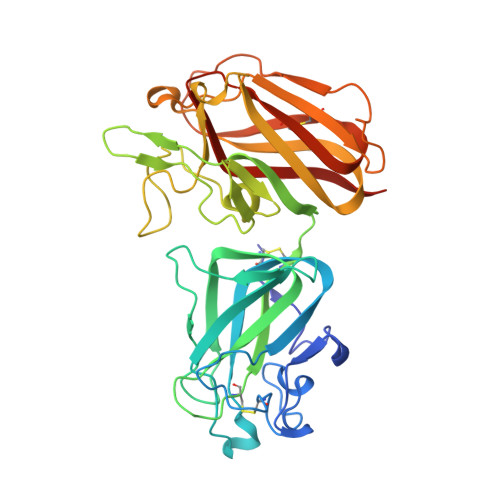
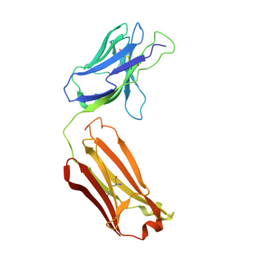
Structure of the Discoidin Domain Receptor 1 Extracellular Region Bound to an Inhibitory Fab Fragment Reveals Features Important for Signaling.

Carafoli, F.Mayer, M.C.Shiraishi, K.Pecheva, M.A.Chan, L.Y.Nan, R.Leitinger, B.Hohenester, E.

(2012) Structure 20: 688

  • DOI: https://doi.org/10.1016/j.str.2012.02.011
  • Primary Citation of Related Structures:  
    4AG4

  • PubMed Abstract: 

    The discoidin domain receptors, DDR1 and DDR2, are constitutively dimeric receptor tyrosine kinases that are activated by triple-helical collagen. Aberrant DDR signaling contributes to several human pathologies, including many cancers. We have generated monoclonal antibodies (mAbs) that inhibit DDR1 signaling without interfering with collagen binding. The crystal structure of the monomeric DDR1 extracellular region bound to the Fab fragment of mAb 3E3 reveals that the collagen-binding discoidin (DS) domain is tightly associated with the following DS-like domain, which contains the epitopes of all mAbs. A conserved surface patch in the DS domain outside the collagen-binding site is shown to be required for signaling. Thus, the active conformation of the DDR1 dimer involves collagen-induced contacts between the DS domains, in addition to the previously identified association of transmembrane helices. The mAbs likely inhibit signaling by sterically blocking the extracellular association of DDR1 subunits.


  • Organizational Affiliation

    Department of Life Sciences, Imperial College London, London SW7 2AZ, UK.


Macromolecules
Find similar proteins by:  (by identity cutoff)  |  3D Structure
Entity ID: 1
MoleculeChains Sequence LengthOrganismDetailsImage
EPITHELIAL DISCOIDIN DOMAIN-CONTAINING RECEPTOR 1351Homo sapiensMutation(s): 0 
EC: 2.7.10.1
UniProt & NIH Common Fund Data Resources
Find proteins for Q08345 (Homo sapiens)
Explore Q08345 
Go to UniProtKB:  Q08345
PHAROS:  Q08345
GTEx:  ENSG00000204580 
Entity Groups  
Sequence Clusters30% Identity50% Identity70% Identity90% Identity95% Identity100% Identity
UniProt GroupQ08345
Sequence Annotations
Expand
  • Reference Sequence
Find similar proteins by:  (by identity cutoff)  |  3D Structure
Entity ID: 2
MoleculeChains Sequence LengthOrganismDetailsImage
MONOCLONAL ANTIBODY 3E3 HEAVY CHAINB [auth H]215Mus musculusMutation(s): 0 
Entity Groups  
Sequence Clusters30% Identity50% Identity70% Identity90% Identity95% Identity100% Identity
Sequence Annotations
Expand
  • Reference Sequence
Find similar proteins by:  (by identity cutoff)  |  3D Structure
Entity ID: 3
MoleculeChains Sequence LengthOrganismDetailsImage
MONOCLONAL ANTIBODY 3E3 LIGHT CHAINC [auth L]213Mus musculusMutation(s): 0 
Entity Groups  
Sequence Clusters30% Identity50% Identity70% Identity90% Identity95% Identity100% Identity
Sequence Annotations
Expand
  • Reference Sequence
Oligosaccharides

Help

Entity ID: 4
MoleculeChains Length2D Diagram Glycosylation3D Interactions
2-acetamido-2-deoxy-beta-D-glucopyranose-(1-4)-2-acetamido-2-deoxy-beta-D-glucopyranoseD [auth B],
E [auth C]
2N-Glycosylation
Glycosylation Resources
GlyTouCan:  G42666HT
GlyCosmos:  G42666HT
GlyGen:  G42666HT
Experimental Data & Validation

Experimental Data

  • Method: X-RAY DIFFRACTION
  • Resolution: 2.80 Å
  • R-Value Free: 0.285 
  • R-Value Work: 0.216 
  • R-Value Observed: 0.216 
  • Space Group: C 2 2 21
Unit Cell:
Length ( Å )Angle ( ˚ )
a = 102.51α = 90
b = 251.48β = 90
c = 75.37γ = 90
Software Package:
Software NamePurpose
CNSrefinement
MOSFLMdata reduction
SCALAdata scaling
PHASERphasing

Structure Validation

View Full Validation Report


Entry History 

Deposition Data

Revision History  (Full details and data files)

  • Version 1.0: 2012-04-18
    Type: Initial release
  • Version 2.0: 2020-07-29
    Type: Remediation
    Reason: Carbohydrate remediation
    Changes: Atomic model, Data collection, Derived calculations, Other, Structure summary
  • Version 2.1: 2023-12-20
    Changes: Data collection, Database references, Refinement description, Structure summary