4A3Q

The 2.15 Angstrom resolution crystal structure of Staphylococcus aureus alanine racemase


Experimental Data Snapshot

  • Method: X-RAY DIFFRACTION
  • Resolution: 2.15 Å
  • R-Value Free: 0.236 
  • R-Value Work: 0.188 
  • R-Value Observed: 0.191 

wwPDB Validation   3D Report Full Report


Ligand Structure Quality Assessment 


This is version 1.2 of the entry. See complete history


Literature

Structural Features and Kinetic Characterization of Alanine Racemase from Staphylococcus Aureus (Mu50)

Scaletti, E.R.Luckner, S.R.Krause, K.L.

(2012) Acta Crystallogr D Biol Crystallogr 68: 82

  • DOI: https://doi.org/10.1107/S0907444911050682
  • Primary Citation of Related Structures:  
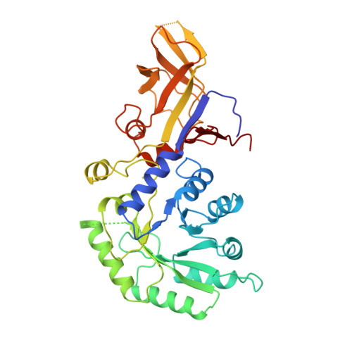
    4A3Q

  • PubMed Abstract: 

    Staphylococcus aureus is an opportunistic Gram-positive bacterium which causes a wide variety of diseases ranging from minor skin infections to potentially fatal conditions such as pneumonia, meningitis and septicaemia. The pathogen is a leading cause of nosocomial acquired infections, a problem that is exacerbated by the existence of methicillin- and glycopeptide antibiotic-resistant strains which can be challenging to treat. Alanine racemase (Alr) is a pyridoxal-5'-phosphate-dependent enzyme which catalyzes reversible racemization between enantiomers of alanine. As D-alanine is an essential component of the bacterial cell-wall peptidoglycan, inhibition of Alr is lethal to prokaryotes. Additionally, while ubiquitous amongst bacteria, this enzyme is absent in humans and most eukaryotes, making it an excellent antibiotic drug target. The crystal structure of S. aureus alanine racemase (Alr(Sas)), the sequence of which corresponds to that from the highly antibiotic-resistant Mu50 strain, has been solved to 2.15 Å resolution. Comparison of the Alr(Sas) structure with those of various alanine racemases demonstrates a conserved overall fold, with the enzyme sharing most similarity to those from other Gram-positive bacteria. Structural examination indicates that the active-site binding pocket, dimer interface and active-site entryway of the enzyme are potential targets for structure-aided inhibitor design. Kinetic constants were calculated in this study and are reported here. The potential for a disulfide bond in this structure is noted. This structural and biochemical information provides a template for future structure-based drug-development efforts targeting Alr(Sas).


  • Organizational Affiliation

    Department of Biochemistry, University of Otago, Dunedin, New Zealand.


Macromolecules
Find similar proteins by:  (by identity cutoff)  |  3D Structure
Entity ID: 1
MoleculeChains Sequence LengthOrganismDetailsImage
ALANINE RACEMASE 1
A, B
382Staphylococcus aureusMutation(s): 0 
EC: 5.1.1.1
UniProt
Find proteins for P63479 (Staphylococcus aureus (strain Mu50 / ATCC 700699))
Explore P63479 
Go to UniProtKB:  P63479
Entity Groups  
Sequence Clusters30% Identity50% Identity70% Identity90% Identity95% Identity100% Identity
UniProt GroupP63479
Sequence Annotations
Expand
  • Reference Sequence
Small Molecules
Ligands 3 Unique
IDChains Name / Formula / InChI Key2D Diagram3D Interactions
PLP
Query on PLP

Download Ideal Coordinates CCD File 
C [auth A],
K [auth B]
PYRIDOXAL-5'-PHOSPHATE
C8 H10 N O6 P
NGVDGCNFYWLIFO-UHFFFAOYSA-N
SO4
Query on SO4

Download Ideal Coordinates CCD File 
D [auth A]
E [auth A]
F [auth A]
G [auth A]
H [auth A]
D [auth A],
E [auth A],
F [auth A],
G [auth A],
H [auth A],
I [auth A],
L [auth B],
M [auth B],
N [auth B],
O [auth B],
P [auth B],
Q [auth B],
R [auth B]
SULFATE ION
O4 S
QAOWNCQODCNURD-UHFFFAOYSA-L
ACT
Query on ACT

Download Ideal Coordinates CCD File 
J [auth A],
S [auth B]
ACETATE ION
C2 H3 O2
QTBSBXVTEAMEQO-UHFFFAOYSA-M
Experimental Data & Validation

Experimental Data

  • Method: X-RAY DIFFRACTION
  • Resolution: 2.15 Å
  • R-Value Free: 0.236 
  • R-Value Work: 0.188 
  • R-Value Observed: 0.191 
  • Space Group: P 21 21 21
Unit Cell:
Length ( Å )Angle ( ˚ )
a = 65.14α = 90
b = 113.94β = 90
c = 125.99γ = 90
Software Package:
Software NamePurpose
REFMACrefinement
MOSFLMdata reduction
SCALAdata scaling
PHASERphasing

Structure Validation

View Full Validation Report


Ligand Structure Quality Assessment 


Entry History 

Deposition Data

Revision History  (Full details and data files)

  • Version 1.0: 2011-12-28
    Type: Initial release
  • Version 1.1: 2012-03-28
    Changes: Other
  • Version 1.2: 2023-12-20
    Changes: Data collection, Database references, Derived calculations, Other, Refinement description