3ZZH

Crystal structure of the amino acid kinase domain from Saccharomyces cerevisiae acetylglutamate kinase in complex with its feed- back inhibitor L-arginine


Experimental Data Snapshot

  • Method: X-RAY DIFFRACTION
  • Resolution: 2.10 Å
  • R-Value Free: 0.215 
  • R-Value Work: 0.180 
  • R-Value Observed: 0.182 

wwPDB Validation   3D Report Full Report


Ligand Structure Quality Assessment 


This is version 1.2 of the entry. See complete history


Literature

Insight on an Arginine Synthesis Metabolon from the Tetrameric Structure of Yeast Acetylglutamate Kinase

De Cima, S.Gil-Ortiz, F.Crabeel, M.Fita, I.Rubio, V.

(2012) PLoS One 7: 34734

  • DOI: https://doi.org/10.1371/journal.pone.0034734
  • Primary Citation of Related Structures:  
    3ZZF, 3ZZG, 3ZZH, 3ZZI, 4AB7

  • PubMed Abstract: 

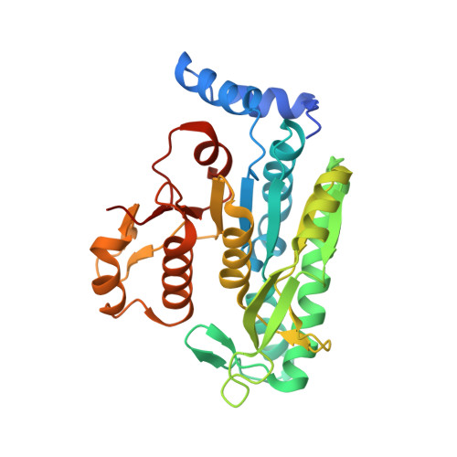
    N-acetyl-L-glutamate kinase (NAGK) catalyzes the second, generally controlling, step of arginine biosynthesis. In yeasts, NAGK exists either alone or forming a metabolon with N-acetyl-L-glutamate synthase (NAGS), which catalyzes the first step and exists only within the metabolon. Yeast NAGK (yNAGK) has, in addition to the amino acid kinase (AAK) domain found in other NAGKs, a ~150-residue C-terminal domain of unclear significance belonging to the DUF619 domain family. We deleted this domain, proving that it stabilizes yNAGK, slows catalysis and modulates feed-back inhibition by arginine. We determined the crystal structures of both the DUF619 domain-lacking yNAGK, ligand-free as well as complexed with acetylglutamate or acetylglutamate and arginine, and of complete mature yNAGK. While all other known arginine-inhibitable NAGKs are doughnut-like hexameric trimers of dimers of AAK domains, yNAGK has as central structure a flat tetramer formed by two dimers of AAK domains. These dimers differ from canonical AAK dimers in the -110° rotation of one subunit with respect to the other. In the hexameric enzymes, an N-terminal extension, found in all arginine-inhibitable NAGKs, forms a protruding helix that interlaces the dimers. In yNAGK, however, it conforms a two-helix platform that mediates interdimeric interactions. Arginine appears to freeze an open inactive AAK domain conformation. In the complete yNAGK structure, two pairs of DUF619 domains flank the AAK domain tetramer, providing a mechanism for the DUF619 domain modulatory functions. The DUF619 domain exhibits the histone acetyltransferase fold, resembling the catalytic domain of bacterial NAGS. However, the putative acetyl CoA site is blocked, explaining the lack of NAGS activity of yNAGK. We conclude that the tetrameric architecture is an adaptation to metabolon formation and propose an organization for this metabolon, suggesting that yNAGK may be a good model also for yeast and human NAGSs.


  • Organizational Affiliation

    Instituto de Biomedicina de Valencia del Consejo Superior de Investigaciones Científicas, Centro de Investigación Biomédica en Red de Enfermedades Raras-ISCIII, Valencia, Spain.


Macromolecules
Find similar proteins by:  (by identity cutoff)  |  3D Structure
Entity ID: 1
MoleculeChains Sequence LengthOrganismDetailsImage
ACETYLGLUTAMATE KINASE
A, B, C, D
307Saccharomyces cerevisiaeMutation(s): 0 
EC: 2.7.2.8
UniProt
Find proteins for Q01217 (Saccharomyces cerevisiae (strain ATCC 204508 / S288c))
Explore Q01217 
Go to UniProtKB:  Q01217
Entity Groups  
Sequence Clusters30% Identity50% Identity70% Identity90% Identity95% Identity100% Identity
UniProt GroupQ01217
Sequence Annotations
Expand
  • Reference Sequence
Small Molecules
Ligands 6 Unique
IDChains Name / Formula / InChI Key2D Diagram3D Interactions
NLG
Query on NLG

Download Ideal Coordinates CCD File 
FA [auth B],
KB [auth D],
M [auth A],
RA [auth C]
N-ACETYL-L-GLUTAMATE
C7 H11 N O5
RFMMMVDNIPUKGG-YFKPBYRVSA-N
ARG
Query on ARG

Download Ideal Coordinates CCD File 
GA [auth C]ARGININE
C6 H15 N4 O2
ODKSFYDXXFIFQN-BYPYZUCNSA-O
MLI
Query on MLI

Download Ideal Coordinates CCD File 
EA [auth B],
JB [auth D],
L [auth A]
MALONATE ION
C3 H2 O4
OFOBLEOULBTSOW-UHFFFAOYSA-L
GOL
Query on GOL

Download Ideal Coordinates CCD File 
PA [auth C]GLYCEROL
C3 H8 O3
PEDCQBHIVMGVHV-UHFFFAOYSA-N
EDO
Query on EDO

Download Ideal Coordinates CCD File 
AA [auth B]
AB [auth D]
BA [auth B]
BB [auth D]
CA [auth B]
AA [auth B],
AB [auth D],
BA [auth B],
BB [auth D],
CA [auth B],
CB [auth D],
DB [auth D],
E [auth A],
EB [auth D],
F [auth A],
FB [auth D],
G [auth A],
GB [auth D],
H [auth A],
HA [auth C],
I [auth A],
IA [auth C],
J [auth A],
JA [auth C],
KA [auth C],
LA [auth C],
MA [auth C],
N [auth B],
NA [auth C],
O [auth B],
OA [auth C],
P [auth B],
Q [auth B],
R [auth B],
S [auth B],
SA [auth D],
T [auth B],
TA [auth D],
U [auth B],
UA [auth D],
V [auth B],
VA [auth D],
W [auth B],
WA [auth D],
X [auth B],
XA [auth D],
Y [auth B],
YA [auth D],
Z [auth B],
ZA [auth D]
1,2-ETHANEDIOL
C2 H6 O2
LYCAIKOWRPUZTN-UHFFFAOYSA-N
CL
Query on CL

Download Ideal Coordinates CCD File 
DA [auth B],
HB [auth D],
IB [auth D],
K [auth A],
QA [auth C]
CHLORIDE ION
Cl
VEXZGXHMUGYJMC-UHFFFAOYSA-M
Binding Affinity Annotations 
IDSourceBinding Affinity
ARG PDBBind:  3ZZH IC50: 4.40e+5 (nM) from 1 assay(s)
Experimental Data & Validation

Experimental Data

  • Method: X-RAY DIFFRACTION
  • Resolution: 2.10 Å
  • R-Value Free: 0.215 
  • R-Value Work: 0.180 
  • R-Value Observed: 0.182 
  • Space Group: P 21 21 21
Unit Cell:
Length ( Å )Angle ( ˚ )
a = 69.489α = 90
b = 99.596β = 90
c = 189.238γ = 90
Software Package:
Software NamePurpose
REFMACrefinement
XDSdata reduction
SCALAdata scaling
MOLREPphasing

Structure Validation

View Full Validation Report


Ligand Structure Quality Assessment 


Entry History 

Deposition Data

Revision History  (Full details and data files)

  • Version 1.0: 2012-05-02
    Type: Initial release
  • Version 1.1: 2012-05-09
    Changes: Other
  • Version 1.2: 2023-12-20
    Changes: Data collection, Database references, Derived calculations, Other, Refinement description