


3ZYR
Structure of the lectin from Platypodium elegans in complex with heptasaccharide
- PDB DOI: https://doi.org/10.2210/pdb3ZYR/pdb
- Classification: SUGAR BINDING PROTEIN
- Organism(s): Platypodium elegans
- Expression System: Escherichia coli BL21(DE3)
- Mutation(s): No
- Deposited: 2011-08-24 Released: 2012-01-11
Experimental Data Snapshot
- Method: X-RAY DIFFRACTION
- Resolution: 1.65 Å
- R-Value Free: 0.188
- R-Value Work: 0.156
- R-Value Observed: 0.157
wwPDB Validation 3D Report Full Report

This is version 2.0 of the entry. See complete history.

Macromolecules
Find similar proteins by:
(by identity cutoff) | 3D Structure
Entity ID: 1 | |||||
|---|---|---|---|---|---|
| Molecule | Chains | Sequence Length | Organism | Details | Image |
| LECTIN | 261 | Platypodium elegans | Mutation(s): 0 | 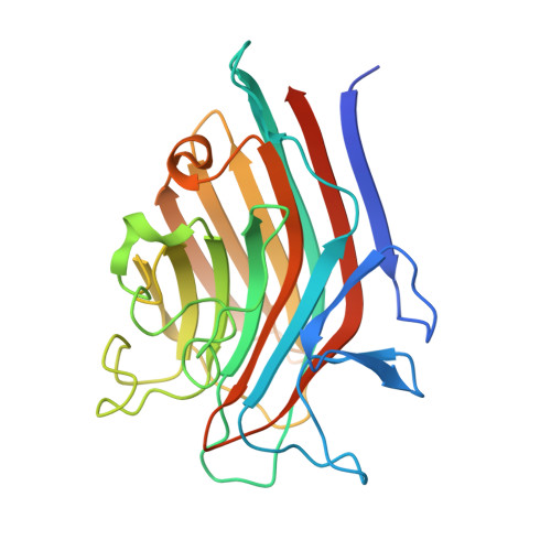 | |
UniProt | |||||
Find proteins for G1EUI6 (Platypodium elegans) Explore G1EUI6 Go to UniProtKB: G1EUI6 | |||||
Entity Groups | |||||
| Sequence Clusters | 30% Identity50% Identity70% Identity90% Identity95% Identity100% Identity | ||||
| UniProt Group | G1EUI6 | ||||
Sequence AnnotationsExpand | |||||
| |||||
Oligosaccharides
Entity ID: 2 | |||||
|---|---|---|---|---|---|
| Molecule | Chains | Length | 2D Diagram | Glycosylation | 3D Interactions |
| 2-acetamido-2-deoxy-beta-D-glucopyranose-(1-2)-alpha-D-mannopyranose-(1-3)-[2-acetamido-2-deoxy-beta-D-glucopyranose-(1-2)-alpha-D-mannopyranose-(1-6)]beta-D-mannopyranose-(1-4)-2-acetamido-2-deoxy-beta-D-glucopyranose-(1-4)-2-acetamido-2-deoxy-beta-D-glucopyranose | C, D | 7 |  | N-Glycosylation | |
Glycosylation Resources | |||||
GlyTouCan: G39213VZ GlyCosmos: G39213VZ GlyGen: G39213VZ | |||||
Small Molecules
| Ligands 5 Unique | |||||
|---|---|---|---|---|---|
| ID | Chains | Name / Formula / InChI Key | 2D Diagram | 3D Interactions | |
| ASN Query on ASN | E [auth A] | ASPARAGINE C4 H8 N2 O3 DCXYFEDJOCDNAF-REOHCLBHSA-N |  | ||
| GOL Query on GOL | H [auth A], J [auth A], K [auth A], N [auth B] | GLYCEROL C3 H8 O3 PEDCQBHIVMGVHV-UHFFFAOYSA-N |  | ||
| URE Query on URE | I [auth A], O [auth B] | UREA C H4 N2 O XSQUKJJJFZCRTK-UHFFFAOYSA-N |  | ||
| MN Query on MN | F [auth A], L [auth B] | MANGANESE (II) ION Mn WAEMQWOKJMHJLA-UHFFFAOYSA-N |  | ||
| CA Query on CA | G [auth A], M [auth B] | CALCIUM ION Ca BHPQYMZQTOCNFJ-UHFFFAOYSA-N |  | ||
Experimental Data & Validation
Experimental Data
- Method: X-RAY DIFFRACTION
- Resolution: 1.65 Å
- R-Value Free: 0.188
- R-Value Work: 0.156
- R-Value Observed: 0.157
- Space Group: P 21 21 21
Unit Cell:
| Length ( Å ) | Angle ( ˚ ) |
|---|---|
| a = 51.4 | α = 90 |
| b = 76.98 | β = 90 |
| c = 125.59 | γ = 90 |
| Software Name | Purpose |
|---|---|
| REFMAC | refinement |
| MOSFLM | data reduction |
| SCALA | data scaling |
| PHASER | phasing |
Entry History
Deposition Data
- Released Date: 2012-01-11 Deposition Author(s): Benevides, R.G., Ganne, G., Cavazza, B.S., Varrot, A., Imberty, A.
Revision History (Full details and data files)
- Version 1.0: 2012-01-11
Type: Initial release - Version 1.1: 2012-01-25
Changes: Other - Version 1.2: 2012-07-25
Changes: Database references, Structure summary - Version 1.3: 2012-08-08
Changes: Database references - Version 2.0: 2020-07-29
Type: Remediation
Reason: Carbohydrate remediation
Changes: Advisory, Atomic model, Data collection, Derived calculations, Other, Structure summary
