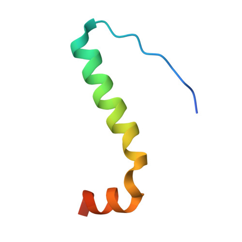
Structure and Kinetic Stability of the P63 Tetramerization Domain.
Natan, E., Joerger, A.C.(2012) J Mol Biol 415: 503
- PubMed: 22100306
- DOI: https://doi.org/10.1016/j.jmb.2011.11.007
- Primary Citation of Related Structures:
3ZY0, 3ZY1 - PubMed Abstract:
The p53 family of transcription factors--comprising p53, p63 and p73--plays an important role in tumor prevention and development. Essential to their function is the formation of tetramers, allowing cooperative binding to their DNA response elements. We solved crystal structures of the human p63 tetramerization domain, showing that p63 forms a dimer of dimers with D₂ symmetry composed of highly intertwined monomers. The primary dimers are formed via an intramolecular β-sheet and hydrophobic helix packing (H1), a hallmark of all p53 family members. Like p73, but unlike p53, p63 requires a second helix (H2) to stabilize the architecture of the tetramer. In order to investigate the impact of structural differences on tetramer stability, we measured the subunit exchange reaction of p53 family homotetramers by nanoflow electrospray mass spectrometry. There were differences in both the kinetics and the pattern of the exchange reaction, with the p53 and p63 tetramers exhibiting much faster exchange kinetics than p73. The structural similarity between p63 and p73 rationalizes previous observations that p63 and p73 form mixed tetramers, and the kinetic data reveal the dissociation of the p73 homotetramers as the rate-limiting step for heterotetramer formation. Differential stability of the tetramers may play an important role in the cross talk between different isoforms and regulation of p53, p63 and p73 function in the cell cycle.
Organizational Affiliation:
Medical Research Council Laboratory of Molecular Biology, Hills Road, Cambridge CB2 0QH, UK.