3ZW6

MODEL OF HEXAMERIC AAA DOMAIN ARRANGEMENT OF GREEN-TYPE RUBISCO ACTIVASE FROM TOBACCO.


Experimental Data Snapshot

  • Method: ELECTRON MICROSCOPY
  • Resolution: 20.0 Å
  • Aggregation State: PARTICLE 
  • Reconstruction Method: SINGLE PARTICLE 

wwPDB Validation   3D Report Full Report


This is version 1.4 of the entry. See complete history


Literature

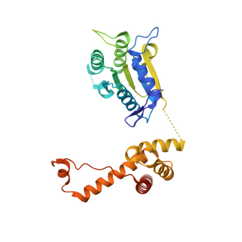
Structure of Green-Type Rubisco Activase from Tobacco

Stotz, M.Mueller-Cajar, O.Ciniawsky, S.Wendler, P.Hartl, F.U.Bracher, A.Hayer-Hartl, M.

(2011) Nat Struct Mol Biol 18: 1366

  • DOI: https://doi.org/10.1038/nsmb.2171
  • Primary Citation of Related Structures:  
    3T15, 3ZW6

  • PubMed Abstract: 

    Rubisco, the enzyme that catalyzes the fixation of atmospheric CO(2) in photosynthesis, is subject to inactivation by inhibitory sugar phosphates. Here we report the 2.95-Å crystal structure of Nicotiana tabacum Rubisco activase (Rca), the enzyme that facilitates the removal of these inhibitors. Rca from tobacco has a classical AAA(+)-protein domain architecture. Although Rca populates a range of oligomeric states when in solution, it forms a helical arrangement with six subunits per turn when in the crystal. However, negative-stain electron microscopy of the active mutant R294V suggests that Rca functions as a hexamer. The residues determining species specificity for Rubisco are located in a helical insertion of the C-terminal domain and probably function in conjunction with the N-domain in Rubisco recognition. Loop segments exposed toward the central pore of the hexamer are required for the ATP-dependent remodeling of Rubisco, resulting in the release of inhibitory sugar.


  • Organizational Affiliation

    Department of Cellular Biochemistry, Max Planck Institute of Biochemistry, Martinsried, Germany.


Macromolecules
Find similar proteins by:  (by identity cutoff)  |  3D Structure
Entity ID: 1
MoleculeChains Sequence LengthOrganismDetailsImage
RIBULOSE BISPHOSPHATE CARBOXYLASE/OXYGENASE ACTIVASE 1, CHLOROPLASTIC
A, B, C, D, E
A, B, C, D, E, F
293Nicotiana tabacumMutation(s): 0 
UniProt
Find proteins for Q40460 (Nicotiana tabacum)
Explore Q40460 
Go to UniProtKB:  Q40460
Entity Groups  
Sequence Clusters30% Identity50% Identity70% Identity90% Identity95% Identity100% Identity
UniProt GroupQ40460
Sequence Annotations
Expand
  • Reference Sequence
Experimental Data & Validation

Experimental Data

  • Method: ELECTRON MICROSCOPY
  • Resolution: 20.0 Å
  • Aggregation State: PARTICLE 
  • Reconstruction Method: SINGLE PARTICLE 
EM Software:
TaskSoftware PackageVersion
RECONSTRUCTIONIMAGIC
RECONSTRUCTIONMRC IMAGE PROCESSING PACKAGE
RECONSTRUCTIONSPIDER

Structure Validation

View Full Validation Report


Entry History 

Deposition Data

Revision History  (Full details and data files)

  • Version 1.0: 2011-11-09
    Type: Initial release
  • Version 1.1: 2011-12-14
    Changes: Database references
  • Version 1.2: 2017-08-23
    Changes: Data collection
  • Version 1.3: 2019-10-23
    Changes: Data collection, Derived calculations, Other
  • Version 1.4: 2019-10-30
    Changes: Advisory, Data collection, Derived calculations