3ZVY

PHD finger of human UHRF1 in complex with unmodified histone H3 N- terminal tail


Experimental Data Snapshot

  • Method: X-RAY DIFFRACTION
  • Resolution: 1.95 Å
  • R-Value Free: 0.227 
  • R-Value Work: 0.181 
  • R-Value Observed: 0.184 

wwPDB Validation   3D Report Full Report


This is version 1.2 of the entry. See complete history


Literature

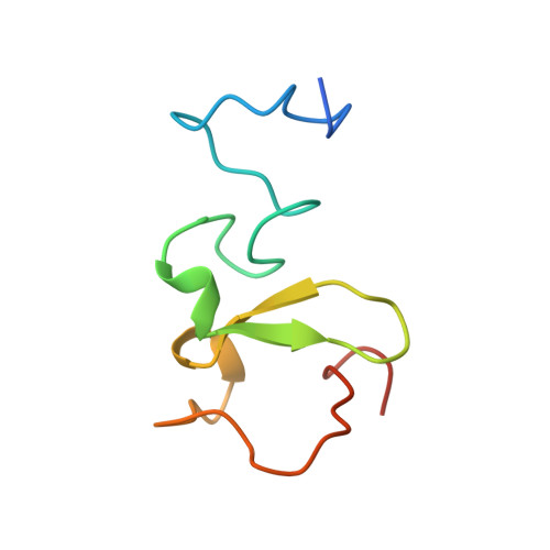
The Phd Finger of Human Uhrf1 Reveals a New Subgroup of Unmethylated Histone H3 Tail Readers.

Lallous, N.Legrand, P.Mcewen, A.G.Ramon-Maiques, S.Samama, J.P.Birck, C.

(2011) PLoS One 6: 27599

  • DOI: https://doi.org/10.1371/journal.pone.0027599
  • Primary Citation of Related Structures:  
    3ZVY, 3ZVZ

  • PubMed Abstract: 

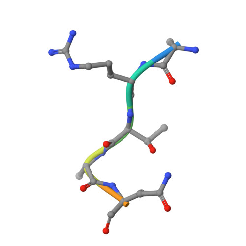
    The human UHRF1 protein (ubiquitin-like containing PHD and RING finger domains 1) has emerged as a potential cancer target due to its implication in cell cycle regulation, maintenance of DNA methylation after replication and heterochromatin formation. UHRF1 functions as an adaptor protein that binds to histones and recruits histone modifying enzymes, like HDAC1 or G9a, which exert their action on chromatin. In this work, we show the binding specificity of the PHD finger of human UHRF1 (huUHRF1-PHD) towards unmodified histone H3 N-terminal tail using native gel electrophoresis and isothermal titration calorimetry. We report the molecular basis of this interaction by determining the crystal structure of huUHRF1-PHD in complex with the histone H3 N-terminal tail. The structure reveals a new mode of histone recognition involving an extra conserved zinc finger preceding the conventional PHD finger region. This additional zinc finger forms part of a large surface cavity that accommodates the side chain of the histone H3 lysine K4 (H3K4) regardless of its methylation state. Mutation of Q330, which specifically interacts with H3K4, to alanine has no effect on the binding, suggesting a loose interaction between huUHRF1-PHD and H3K4. On the other hand, the recognition appears to rely on histone H3R2, which fits snugly into a groove on the protein and makes tight interactions with the conserved aspartates D334 and D337. Indeed, a mutation of the former aspartate disrupts the formation of the complex, while mutating the latter decreases the binding affinity nine-fold.


  • Organizational Affiliation

    Institut National de Santé et de Recherche Médicale U964/Centre National de Recherche Scientifique UMR 7104/Université de Strasbourg, Illkirch, France. nlallous@cnio.es


Macromolecules
Find similar proteins by:  (by identity cutoff)  |  3D Structure
Entity ID: 1
MoleculeChains Sequence LengthOrganismDetailsImage
E3 UBIQUITIN-PROTEIN LIGASE UHRF1
A, B
72Homo sapiensMutation(s): 0 
EC: 6.3.2
UniProt & NIH Common Fund Data Resources
Find proteins for Q96T88 (Homo sapiens)
Explore Q96T88 
Go to UniProtKB:  Q96T88
PHAROS:  Q96T88
GTEx:  ENSG00000276043 
Entity Groups  
Sequence Clusters30% Identity50% Identity70% Identity90% Identity95% Identity100% Identity
UniProt GroupQ96T88
Sequence Annotations
Expand
  • Reference Sequence

Find similar proteins by:  Sequence   |   3D Structure  

Entity ID: 2
MoleculeChains Sequence LengthOrganismDetailsImage
HISTONE H3.1
C, D
8Homo sapiensMutation(s): 0 
UniProt & NIH Common Fund Data Resources
Find proteins for P68431 (Homo sapiens)
Explore P68431 
Go to UniProtKB:  P68431
PHAROS:  P68431
Entity Groups  
UniProt GroupP68431
Sequence Annotations
Expand
  • Reference Sequence
Small Molecules
Ligands 3 Unique
IDChains Name / Formula / InChI Key2D Diagram3D Interactions
TRS
Query on TRS

Download Ideal Coordinates CCD File 
I [auth A],
P [auth B]
2-AMINO-2-HYDROXYMETHYL-PROPANE-1,3-DIOL
C4 H12 N O3
LENZDBCJOHFCAS-UHFFFAOYSA-O
ZN
Query on ZN

Download Ideal Coordinates CCD File 
E [auth A]
F [auth A]
G [auth A]
H [auth A]
J [auth B]
E [auth A],
F [auth A],
G [auth A],
H [auth A],
J [auth B],
K [auth B],
L [auth B],
M [auth B]
ZINC ION
Zn
PTFCDOFLOPIGGS-UHFFFAOYSA-N
CL
Query on CL

Download Ideal Coordinates CCD File 
N [auth B],
O [auth B]
CHLORIDE ION
Cl
VEXZGXHMUGYJMC-UHFFFAOYSA-M
Experimental Data & Validation

Experimental Data

  • Method: X-RAY DIFFRACTION
  • Resolution: 1.95 Å
  • R-Value Free: 0.227 
  • R-Value Work: 0.181 
  • R-Value Observed: 0.184 
  • Space Group: P 43 21 2
Unit Cell:
Length ( Å )Angle ( ˚ )
a = 42.3α = 90
b = 42.3β = 90
c = 182.7γ = 90
Software Package:
Software NamePurpose
PHENIXrefinement
XDSdata reduction
XSCALEdata scaling

Structure Validation

View Full Validation Report


Entry History 

Deposition Data

Revision History  (Full details and data files)

  • Version 1.0: 2011-12-07
    Type: Initial release
  • Version 1.1: 2019-03-06
    Changes: Data collection, Experimental preparation, Other
  • Version 1.2: 2019-05-08
    Changes: Data collection, Experimental preparation