3ZRH

Crystal structure of the Lys29, Lys33-linkage-specific TRABID OTU deubiquitinase domain reveals an Ankyrin-repeat ubiquitin binding domain (AnkUBD)


Experimental Data Snapshot

  • Method: X-RAY DIFFRACTION
  • Resolution: 2.23 Å
  • R-Value Free: 0.248 
  • R-Value Work: 0.200 
  • R-Value Observed: 0.203 

wwPDB Validation   3D Report Full Report


This is version 1.1 of the entry. See complete history


Literature

An Ankyrin-Repeat Ubiquitin-Binding Domain Determines Trabid'S Specificity for Atypical Ubiquitin Chains.

Licchesi, J.D.F.Mieszczanek, J.Mevissen, T.E.T.Rutherford, T.J.Akutsu, M.Virdee, S.Oualid, F.E.Chin, J.W.Ovaa, H.Bienz, M.Komander, D.

(2011) Nat Struct Mol Biol 19: 62

  • DOI: https://doi.org/10.1038/nsmb.2169
  • Primary Citation of Related Structures:  
    3ZRH

  • PubMed Abstract: 

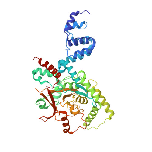
    Eight different types of ubiquitin linkages are present in eukaryotic cells that regulate diverse biological processes. Proteins that mediate specific assembly and disassembly of atypical Lys6, Lys27, Lys29 and Lys33 linkages are mainly unknown. We here reveal how the human ovarian tumor (OTU) domain deubiquitinase (DUB) TRABID specifically hydrolyzes both Lys29- and Lys33-linked diubiquitin. A crystal structure of the extended catalytic domain reveals an unpredicted ankyrin repeat domain that precedes an A20-like catalytic core. NMR analysis identifies the ankyrin domain as a new ubiquitin-binding fold, which we have termed AnkUBD, and DUB assays in vitro and in vivo show that this domain is crucial for TRABID efficiency and linkage specificity. Our data are consistent with AnkUBD functioning as an enzymatic S1' ubiquitin-binding site, which orients a ubiquitin chain so that Lys29 and Lys33 linkages are cleaved preferentially.


  • Organizational Affiliation

    Medical Research Council Laboratory of Molecular Biology, Cambridge, UK.


Macromolecules
Find similar proteins by:  (by identity cutoff)  |  3D Structure
Entity ID: 1
MoleculeChains Sequence LengthOrganismDetailsImage
UBIQUITIN THIOESTERASE ZRANB1454Homo sapiensMutation(s): 0 
EC: 3.4.19.12
UniProt & NIH Common Fund Data Resources
Find proteins for Q9UGI0 (Homo sapiens)
Explore Q9UGI0 
Go to UniProtKB:  Q9UGI0
PHAROS:  Q9UGI0
GTEx:  ENSG00000019995 
Entity Groups  
Sequence Clusters30% Identity50% Identity70% Identity90% Identity95% Identity100% Identity
UniProt GroupQ9UGI0
Sequence Annotations
Expand
  • Reference Sequence
Experimental Data & Validation

Experimental Data

  • Method: X-RAY DIFFRACTION
  • Resolution: 2.23 Å
  • R-Value Free: 0.248 
  • R-Value Work: 0.200 
  • R-Value Observed: 0.203 
  • Space Group: P 21 21 21
Unit Cell:
Length ( Å )Angle ( ˚ )
a = 60.4α = 90
b = 72.15β = 90
c = 133.01γ = 90
Software Package:
Software NamePurpose
PHENIXrefinement
MOSFLMdata reduction
SCALAdata scaling
SHARPphasing

Structure Validation

View Full Validation Report


Entry History 

Deposition Data

Revision History  (Full details and data files)

  • Version 1.0: 2011-12-14
    Type: Initial release
  • Version 1.1: 2012-01-25
    Changes: Other