Structures of Phytophthora Rxlr Effector Proteins: A Conserved But Adaptable Fold Underpins Functional Diversity.
Boutemy, L.S., King, S.R.F., Win, J., Hughes, R.K., Clarke, T.A., Blumenschein, T.M.A., Kamoun, S., Banfield, M.J.(2011) J Biol Chem 286: 35834
- PubMed: 21813644
- DOI: https://doi.org/10.1074/jbc.M111.262303
- Primary Citation of Related Structures:
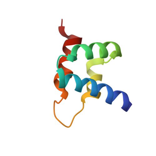
3ZR8, 3ZRG - PubMed Abstract:
Phytopathogens deliver effector proteins inside host plant cells to promote infection. These proteins can also be sensed by the plant immune system, leading to restriction of pathogen growth. Effector genes can display signatures of positive selection and rapid evolution, presumably a consequence of their co-evolutionary arms race with plants. The molecular mechanisms underlying how effectors evolve to gain new virulence functions and/or evade the plant immune system are poorly understood. Here, we report the crystal structures of the effector domains from two oomycete RXLR proteins, Phytophthora capsici AVR3a11 and Phytophthora infestans PexRD2. Despite sharing <20% sequence identity in their effector domains, they display a conserved core α-helical fold. Bioinformatic analyses suggest that the core fold occurs in ∼44% of annotated Phytophthora RXLR effectors, both as a single domain and in tandem repeats of up to 11 units. Functionally important and polymorphic residues map to the surface of the structures, and PexRD2, but not AVR3a11, oligomerizes in planta. We conclude that the core α-helical fold enables functional adaptation of these fast evolving effectors through (i) insertion/deletions in loop regions between α-helices, (ii) extensions to the N and C termini, (iii) amino acid replacements in surface residues, (iv) tandem domain duplications, and (v) oligomerization. We hypothesize that the molecular stability provided by this core fold, combined with considerable potential for plasticity, underlies the evolution of effectors that maintain their virulence activities while evading recognition by the plant immune system.
Organizational Affiliation:
Department of Biological Chemistry, John Innes Centre, Norwich Research Park, Norwich NR4 7UH, United Kingdom.