3ZQ4

Unusual, dual endo- and exo-nuclease activity in the degradosome explained by crystal structure analysis of RNase J1


Experimental Data Snapshot

  • Method: X-RAY DIFFRACTION
  • Resolution: 3.00 Å
  • R-Value Free: 0.258 
  • R-Value Work: 0.214 
  • R-Value Observed: 0.216 

wwPDB Validation   3D Report Full Report


This is version 1.4 of the entry. See complete history


Literature

Unusual, Dual Endo- and Exonuclease Activity in the Degradosome Explained by Crystal Structure Analysis of Rnase J1.

Newman, J.A.Hewitt, L.Rodrigues, C.Solovyova, A.Harwood, C.R.Lewis, R.J.

(2011) Structure 19: 1241

  • DOI: https://doi.org/10.1016/j.str.2011.06.017
  • Primary Citation of Related Structures:  
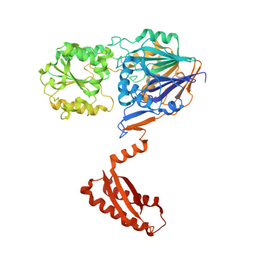
    3ZQ4

  • PubMed Abstract: 

    RNase J is an essential enzyme in Bacillus subtilis with unusual dual endonuclease and 5'-to-3' exonuclease activities that play an important role in the maturation and degradation of mRNA. RNase J is also a component of the recently identified "degradosome" of B. subtilis. We report the crystal structure of RNase J1 from B. subtilis to 3.0 Å resolution, analysis of which reveals it to be in an open conformation suitable for binding substrate RNA. RNase J is a member of the β-CASP family of zinc-dependent metallo-β-lactamases. We have exploited this similarity in constructing a model for an RNase J1:RNA complex. Analysis of this model reveals candidate-stacking interactions with conserved aromatic side chains, providing a molecular basis for the observed enzyme activity. Comparisons of the B. subtilis RNase J structure with related enzymes reveal key differences that provide insights into conformational changes during catalysis and the role of the C-terminal domain.


  • Organizational Affiliation

    Institute for Cell and Molecular Biosciences, Newcastle University, Newcastle-upon-Tyne NE2 4HH, UK.


Macromolecules
Find similar proteins by:  (by identity cutoff)  |  3D Structure
Entity ID: 1
MoleculeChains Sequence LengthOrganismDetailsImage
RIBONUCLEASE J 1A,
B [auth C],
C [auth D],
D [auth E]
555Bacillus subtilis subsp. subtilis str. 168Mutation(s): 0 
EC: 3.1
UniProt
Find proteins for Q45493 (Bacillus subtilis (strain 168))
Explore Q45493 
Go to UniProtKB:  Q45493
Entity Groups  
Sequence Clusters30% Identity50% Identity70% Identity90% Identity95% Identity100% Identity
UniProt GroupQ45493
Sequence Annotations
Expand
  • Reference Sequence
Small Molecules
Ligands 2 Unique
IDChains Name / Formula / InChI Key2D Diagram3D Interactions
ZN
Query on ZN

Download Ideal Coordinates CCD File 
E [auth A]
F [auth A]
H [auth C]
I [auth C]
L [auth D]
E [auth A],
F [auth A],
H [auth C],
I [auth C],
L [auth D],
M [auth D],
N [auth E],
O [auth E]
ZINC ION
Zn
PTFCDOFLOPIGGS-UHFFFAOYSA-N
CA
Query on CA

Download Ideal Coordinates CCD File 
G [auth A],
J [auth C],
K [auth D],
P [auth E]
CALCIUM ION
Ca
BHPQYMZQTOCNFJ-UHFFFAOYSA-N
Experimental Data & Validation

Experimental Data

  • Method: X-RAY DIFFRACTION
  • Resolution: 3.00 Å
  • R-Value Free: 0.258 
  • R-Value Work: 0.214 
  • R-Value Observed: 0.216 
  • Space Group: P 21 21 2
Unit Cell:
Length ( Å )Angle ( ˚ )
a = 177.823α = 90
b = 209.43β = 90
c = 74.106γ = 90
Software Package:
Software NamePurpose
PHENIXrefinement
XDSdata reduction
XDSdata scaling
PHASERphasing

Structure Validation

View Full Validation Report


Entry History 

Deposition Data

Revision History  (Full details and data files)

  • Version 1.0: 2011-09-14
    Type: Initial release
  • Version 1.1: 2018-06-20
    Changes: Advisory, Data collection, Derived calculations, Source and taxonomy
  • Version 1.2: 2019-02-20
    Changes: Advisory, Data collection, Derived calculations
  • Version 1.3: 2019-07-10
    Changes: Data collection
  • Version 1.4: 2023-12-20
    Changes: Data collection, Database references, Derived calculations, Other, Refinement description