3ZLV

Crystal structure of mouse acetylcholinesterase in complex with tabun and HI-6


Experimental Data Snapshot

  • Method: X-RAY DIFFRACTION
  • Resolution: 2.50 Å
  • R-Value Free: 0.226 
  • R-Value Work: 0.176 
  • R-Value Observed: 0.177 

wwPDB Validation   3D Report Full Report


Ligand Structure Quality Assessment 


This is version 1.5 of the entry. See complete history


Literature

Catalytic-Site Conformational Equilibrium in Nerve-Agent Adducts of Acetylcholinesterase; Possible Implications for the Hi-6 Antidote Substrate Specificity.

Artursson, E.Andersson, P.O.Akfur, C.Linusson, A.Borjegren, S.Ekstrom, F.

(2013) Biochem Pharmacol 85: 1389

  • DOI: https://doi.org/10.1016/j.bcp.2013.01.016
  • Primary Citation of Related Structures:  
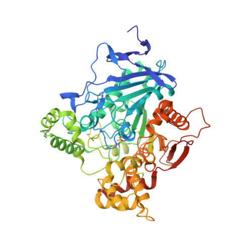
    3ZLU, 3ZLV

  • PubMed Abstract: 

    Nerve agents such as tabun, cyclosarin and Russian VX inhibit the essential enzyme acetylcholinesterase (AChE) by organophosphorylating the catalytic serine residue. Nucleophiles, such as oximes, are used as antidotes as they can reactivate and restore the function of the inhibited enzyme. The oxime HI-6 shows a notably low activity on tabun adducts but can effectively reactivate adducts of cyclosarin and Russian VX. To examine the structural basis for the pronounced substrate specificity of HI-6, we determined the binary crystal structures of Mus musculus AChE (mAChE) conjugated by cyclosarin and Russian VX and found a conformational mobility of the side chains of Phe338 and His447. The interaction between HI-6 and tabun-adducts of AChE were subsequently investigated using a combination of time resolved fluorescence spectroscopy and X-ray crystallography. Our findings show that HI-6 binds to tabun inhibited Homo sapiens AChE (hAChE) with an IC50 value of 300μM and suggest that the reactive nucleophilic moiety of HI-6 is excluded from the phosphorus atom of tabun. We propose that a conformational mobility of the side-chains of Phe338 and His447 is a common feature in nerve-agent adducts of AChE. We also suggest that the conformational mobility allow HI-6 to reactivate conjugates of cyclosarin and Russian VX while a reduced mobility in tabun conjugated AChE results in steric hindrance that prevents efficient reactivation.


  • Organizational Affiliation

    Swedish Defence Research Agency, CBRN, Defence and Security, Umeå, Sweden.


Macromolecules
Find similar proteins by:  (by identity cutoff)  |  3D Structure
Entity ID: 1
MoleculeChains Sequence LengthOrganismDetailsImage
ACETYLCHOLINESTERASE
A, B
543Mus musculusMutation(s): 0 
EC: 3.1.1.7
UniProt & NIH Common Fund Data Resources
Find proteins for P21836 (Mus musculus)
Explore P21836 
Go to UniProtKB:  P21836
IMPC:  MGI:87876
Entity Groups  
Sequence Clusters30% Identity50% Identity70% Identity90% Identity95% Identity100% Identity
UniProt GroupP21836
Sequence Annotations
Expand
  • Reference Sequence
Small Molecules
Ligands 4 Unique
IDChains Name / Formula / InChI Key2D Diagram3D Interactions
PE4
Query on PE4

Download Ideal Coordinates CCD File 
J [auth B]2-{2-[2-(2-{2-[2-(2-ETHOXY-ETHOXY)-ETHOXY]-ETHOXY}-ETHOXY)-ETHOXY]-ETHOXY}-ETHANOL
C16 H34 O8
PJWQOENWHPEPKI-UHFFFAOYSA-N
HI6
Query on HI6

Download Ideal Coordinates CCD File 
C [auth A],
H [auth B]
4-(AMINOCARBONYL)-1-[({2-[(E)-(HYDROXYIMINO)METHYL]PYRIDINIUM-1-YL}METHOXY)METHYL]PYRIDINIUM
C14 H16 N4 O3
FJZDLOMCEPUCII-UHFFFAOYSA-P
1KA
Query on 1KA

Download Ideal Coordinates CCD File 
E [auth A],
F [auth A],
G [auth A],
I [auth B],
K [auth B]
(2-hydroxyethoxy)acetaldehyde
C4 H8 O3
DSGGHBUAHUMMHN-UHFFFAOYSA-N
CO3
Query on CO3

Download Ideal Coordinates CCD File 
D [auth A]CARBONATE ION
C O3
BVKZGUZCCUSVTD-UHFFFAOYSA-L
Modified Residues  1 Unique
IDChains TypeFormula2D DiagramParent
SUN
Query on SUN
A, B
L-PEPTIDE LINKINGC7 H17 N2 O5 PSER
Binding Affinity Annotations 
IDSourceBinding Affinity
HI6 BindingDB:  3ZLV Kd: min: 1.50e+4, max: 2.34e+5 (nM) from 6 assay(s)
IC50: min: 6.36e+5, max: 1.36e+6 (nM) from 3 assay(s)
PDBBind:  3ZLV IC50: 3.00e+5 (nM) from 1 assay(s)
Experimental Data & Validation

Experimental Data

  • Method: X-RAY DIFFRACTION
  • Resolution: 2.50 Å
  • R-Value Free: 0.226 
  • R-Value Work: 0.176 
  • R-Value Observed: 0.177 
  • Space Group: P 21 21 21
Unit Cell:
Length ( Å )Angle ( ˚ )
a = 79.57α = 90
b = 112.38β = 90
c = 226.99γ = 90
Software Package:
Software NamePurpose
PHENIXrefinement
XDSdata reduction
SCALAdata scaling
REFMACphasing

Structure Validation

View Full Validation Report


Ligand Structure Quality Assessment 


Entry History 

Deposition Data

Revision History  (Full details and data files)

  • Version 1.0: 2013-03-27
    Type: Initial release
  • Version 1.1: 2013-08-07
    Changes: Database references
  • Version 1.2: 2013-08-14
    Changes: Atomic model
  • Version 1.3: 2015-04-01
    Changes: Data collection
  • Version 1.4: 2018-01-17
    Changes: Data collection
  • Version 1.5: 2023-12-20
    Changes: Data collection, Database references, Derived calculations, Other, Refinement description