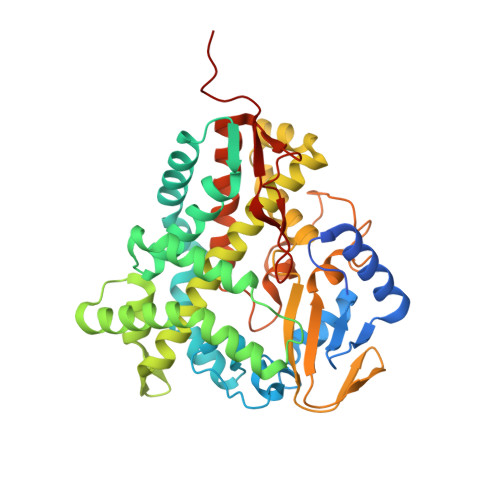
3ZKP

Structure of a mutant of P450 EryK in complex with erythromycin B.


Experimental Data Snapshot

  • Method: X-RAY DIFFRACTION
  • Resolution: 2.00 Å
  • R-Value Free: 0.215 
  • R-Value Work: 0.163 
  • R-Value Observed: 0.166 

wwPDB Validation   3D Report Full Report


Ligand Structure Quality Assessment 


This is version 1.2 of the entry. See complete history


Literature

Redirecting P450 Eryk Specificity by Rational Site-Directed Mutagenesis.

Montemiglio, L.C.Macone, A.Ardiccioni, C.Avella, G.Vallone, B.Savino, C.

(2013) Biochemistry 52: 3678

  • DOI: https://doi.org/10.1021/bi400223j
  • Primary Citation of Related Structures:  
    3ZKP

  • PubMed Abstract: 

    The C-12 hydroxylase EryK is a bacterial cytochrome P450, active during one of the final tailoring steps of erythromycin A (ErA) biosynthesis. Its tight substrate specificity, restricted to the metabolic intermediate ErD, leads to the accumulation in the culture broth of a shunt metabolite, ErB, that originates from the competitive action of a methyltranferase on the substrate of EryK. Although the methylation of the mycarosyl moiety represents the only difference between the two metabolites, EryK exhibits very low conversion of ErB in ErA via a parallel pathway. Given its limited antimicrobial activity and its moderate toxicity, contamination by such a byproduct decreases the yield and purity of the antibiotic. In this study, EryK has been redesigned to make it suitable for industrial application. Taking advantage of the three-dimensional structure of the enzyme in complex with ErD, three single active-site mutants of EryK (M86A, H88E, and E89L) have been designed to allow hydroxylation of the nonphysiological substrate ErB. The binding and catalytic properties of these three variants on both ErD and ErB have been analyzed. Interestingly, we found the mutation of Met 86 to Ala to yield enzymatic activity on both ErB and ErD. The three-dimensional structure of the complex of mutated EryK with ErB revealed that the mutation allows ErB to accommodate in the active site of the enzyme and to induce its closure, thus assuring the progress of the catalytic reaction. Therefore, by single mutation the fine substrate recognition, active site closure, and locking were recovered.


  • Organizational Affiliation

    Istituto Pasteur-Fondazione Cenci Bolognetti and Istituto di Biologia e Patologia Molecolari del CNR, Dipartimento di Scienze Biochimiche "A. Rossi Fanelli", Sapienza Università di Roma , Piazzale A. Moro 5, 00185 Rome, Italy.


Macromolecules
Find similar proteins by:  (by identity cutoff)  |  3D Structure
Entity ID: 1
MoleculeChains Sequence LengthOrganismDetailsImage
ERYTHROMYCIN C-12 HYDROXYLASE411Saccharopolyspora erythraea NRRL 2338Mutation(s): 1 
EC: 1.14.13.154
UniProt
Find proteins for P48635 (Saccharopolyspora erythraea (strain ATCC 11635 / DSM 40517 / JCM 4748 / NBRC 13426 / NCIMB 8594 / NRRL 2338))
Explore P48635 
Go to UniProtKB:  P48635
Entity Groups  
Sequence Clusters30% Identity50% Identity70% Identity90% Identity95% Identity100% Identity
UniProt GroupP48635
Sequence Annotations
Expand
  • Reference Sequence
Small Molecules
Ligands 2 Unique
IDChains Name / Formula / InChI Key2D Diagram3D Interactions
ERB
Query on ERB

Download Ideal Coordinates CCD File 
C [auth A]Erythromycin B
C37 H67 N O12
IDRYSCOQVVUBIJ-PPGFLMPOSA-N
HEM
Query on HEM

Download Ideal Coordinates CCD File 
B [auth A]PROTOPORPHYRIN IX CONTAINING FE
C34 H32 Fe N4 O4
KABFMIBPWCXCRK-RGGAHWMASA-L
Binding Affinity Annotations 
IDSourceBinding Affinity
ERB Binding MOAD:  3ZKP Kd: 1.40e+4 (nM) from 1 assay(s)
Experimental Data & Validation

Experimental Data

  • Method: X-RAY DIFFRACTION
  • Resolution: 2.00 Å
  • R-Value Free: 0.215 
  • R-Value Work: 0.163 
  • R-Value Observed: 0.166 
  • Space Group: P 1 21 1
Unit Cell:
Length ( Å )Angle ( ˚ )
a = 57.753α = 90
b = 36.597β = 92.93
c = 96.273γ = 90
Software Package:
Software NamePurpose
REFMACrefinement
HKL-2000data reduction
SCALEPACKdata scaling
REFMACphasing

Structure Validation

View Full Validation Report


Ligand Structure Quality Assessment 


Entry History 

Deposition Data

Revision History  (Full details and data files)

  • Version 1.0: 2013-05-01
    Type: Initial release
  • Version 1.1: 2013-08-07
    Changes: Database references, Other, Structure summary
  • Version 1.2: 2023-12-20
    Changes: Data collection, Database references, Derived calculations, Other, Refinement description