3ZI0

Structure of Mycobacterium tuberculosis DXR in complex with a fosmidomycin analogue


Experimental Data Snapshot

  • Method: X-RAY DIFFRACTION
  • Resolution: 1.90 Å
  • R-Value Free: 0.231 
  • R-Value Work: 0.205 
  • R-Value Observed: 0.206 

wwPDB Validation   3D Report Full Report


Ligand Structure Quality Assessment 


This is version 1.0 of the entry. See complete history


Literature

Dxr Inhibition by Potent Mono- and Disubstituted Fosmidomycin Analogues.

Jansson, A.M.Wieckowska, A.Bjorkelid, C.Yahiaoui, S.Sooriyaarachchi, S.Lindh, M.Bergfors, T.Dharavath, S.Desroses, M.Suresh, S.Andaloussi, M.Nikhil, R.Sreevalli, S.Srinivasa, B.R.Larhed, M.Jones, T.A.Karlen, A.Mowbray, S.L.

(2013) J Med Chem 56: 6190

  • DOI: https://doi.org/10.1021/jm4006498

  • PubMed Abstract: 

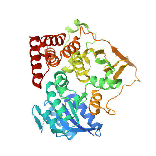
    The antimalarial compound fosmidomycin targets DXR, the enzyme that catalyzes the first committed step in the MEP pathway, producing the essential isoprenoid precursors, isopentenyl diphosphate and dimethylallyl diphosphate. The MEP pathway is used by a number of pathogens, including Mycobacterium tuberculosis and apicomplexan parasites, and differs from the classical mevalonate pathway that is essential in humans. Using a structure-based approach, we designed a number of analogues of fosmidomycin, including a series that are substituted in both the Cα and the hydroxamate positions. The latter proved to be a stable framework for the design of inhibitors that extend from the polar and cramped (and so not easily druggable) substrate-binding site and can, for the first time, bridge the substrate and cofactor binding sites. A number of these compounds are more potent than fosmidomycin in terms of killing Plasmodium falciparum in an in vitro assay; the best has an IC50 of 40 nM.


  • Organizational Affiliation

    Department of Cell and Molecular Biology, Uppsala University , Biomedical Center, Box 596, SE-751 24 Uppsala, Sweden.


Macromolecules
Find similar proteins by:  (by identity cutoff)  |  3D Structure
Entity ID: 1
MoleculeChains Sequence LengthOrganismDetailsImage
1-DEOXY-D-XYLULOSE 5-PHOSPHATE REDUCTOISOMERASE
A, B
397Mycobacterium tuberculosis H37RvMutation(s): 0 
EC: 1.1.1.267
UniProt
Find proteins for P9WNS1 (Mycobacterium tuberculosis (strain ATCC 25618 / H37Rv))
Explore P9WNS1 
Go to UniProtKB:  P9WNS1
Entity Groups  
Sequence Clusters30% Identity50% Identity70% Identity90% Identity95% Identity100% Identity
UniProt GroupP9WNS1
Sequence Annotations
Expand
  • Reference Sequence
Small Molecules
Ligands 2 Unique
IDChains Name / Formula / InChI Key2D Diagram3D Interactions
FM8
Query on FM8

Download Ideal Coordinates CCD File 
C [auth A][(1S)-1-(3,4-dichlorophenyl)-3-{hydroxy[2-(1H-1,2,4-triazol-1-ylmethyl)benzoyl]amino}propyl]phosphonic acid
C19 H19 Cl2 N4 O5 P
CGKYBZFXVQNZBL-SFHVURJKSA-N
MN
Query on MN

Download Ideal Coordinates CCD File 
D [auth B]MANGANESE (II) ION
Mn
WAEMQWOKJMHJLA-UHFFFAOYSA-N
Binding Affinity Annotations 
IDSourceBinding Affinity
FM8 PDBBind:  3ZI0 Kd: 40 (nM) from 1 assay(s)
BindingDB:  3ZI0 Kd: 40 (nM) from 1 assay(s)
IC50: 1.30e+4 (nM) from 1 assay(s)
Binding MOAD:  3ZI0 Kd: 40 (nM) from 1 assay(s)
Experimental Data & Validation

Experimental Data

  • Method: X-RAY DIFFRACTION
  • Resolution: 1.90 Å
  • R-Value Free: 0.231 
  • R-Value Work: 0.205 
  • R-Value Observed: 0.206 
  • Space Group: P 1 21 1
Unit Cell:
Length ( Å )Angle ( ˚ )
a = 67.447α = 90
b = 68.707β = 107.34
c = 85.7γ = 90
Software Package:
Software NamePurpose
REFMACrefinement
MOSFLMdata reduction
SCALAdata scaling
PHASERphasing

Structure Validation

View Full Validation Report


Ligand Structure Quality Assessment 


Entry History 

Deposition Data

Revision History  (Full details and data files)

  • Version 1.0: 2013-10-09
    Type: Initial release