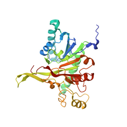
3ZF8

Crystal structure of Saccharomyces cerevisiae Mnn9 in complex with GDP and Mn.


Experimental Data Snapshot

  • Method: X-RAY DIFFRACTION
  • Resolution: 1.98 Å
  • R-Value Free: 0.239 
  • R-Value Work: 0.190 
  • R-Value Observed: 0.191 

wwPDB Validation   3D Report Full Report


Ligand Structure Quality Assessment 


This is version 1.0 of the entry. See complete history


Literature

Yeast Mnn9 is Both a Priming Glycosyltransferase and an Allosteric Activator of Mannan Biosynthesis.

Striebeck, A.Robinson, D.A.Schuettelkopf, A.W.Van Aalten, D.M.F.

(2013) Open Biol 3: 30022

  • DOI: https://doi.org/10.1098/rsob.130022
  • Primary Citation of Related Structures:  
    3ZF8

  • PubMed Abstract: 

    The fungal cell possesses an essential carbohydrate cell wall. The outer layer, mannan, is formed by mannoproteins carrying highly mannosylated O- and N-linked glycans. Yeast mannan biosynthesis is initiated by a Golgi-located complex (M-Pol I) of two GT-62 mannosyltransferases, Mnn9p and Van1p, that are conserved in fungal pathogens. Saccharomyces cerevisiae and Candida albicans mnn9 knockouts show an aberrant cell wall and increased antibiotic sensitivity, suggesting the enzyme is a potential drug target. Here, we present the structure of ScMnn9 in complex with GDP and Mn(2+), defining the fold and catalytic machinery of the GT-62 family. Compared with distantly related GT-78/GT-15 enzymes, ScMnn9 carries an unusual extension. Using a novel enzyme assay and site-directed mutagenesis, we identify conserved amino acids essential for ScMnn9 'priming' α-1,6-mannosyltransferase activity. Strikingly, both the presence of the ScMnn9 protein and its product, but not ScMnn9 catalytic activity, are required to activate subsequent ScVan1 processive α-1,6-mannosyltransferase activity in the M-Pol I complex. These results reveal the molecular basis of mannan synthesis and will aid development of inhibitors targeting this process.


  • Organizational Affiliation

    Division of Molecular Microbiology, University of Dundee, Dundee DD1 5EH, UK.


Macromolecules
Find similar proteins by:  (by identity cutoff)  |  3D Structure
Entity ID: 1
MoleculeChains Sequence LengthOrganismDetailsImage
MANNAN POLYMERASE COMPLEXES SUBUNIT MNN9305Saccharomyces cerevisiae S288CMutation(s): 1 
UniProt
Find proteins for P39107 (Saccharomyces cerevisiae (strain ATCC 204508 / S288c))
Explore P39107 
Go to UniProtKB:  P39107
Entity Groups  
Sequence Clusters30% Identity50% Identity70% Identity90% Identity95% Identity100% Identity
UniProt GroupP39107
Sequence Annotations
Expand
  • Reference Sequence
Small Molecules
Ligands 2 Unique
IDChains Name / Formula / InChI Key2D Diagram3D Interactions
GDP
Query on GDP

Download Ideal Coordinates CCD File 
B [auth A]GUANOSINE-5'-DIPHOSPHATE
C10 H15 N5 O11 P2
QGWNDRXFNXRZMB-UUOKFMHZSA-N
MN
Query on MN

Download Ideal Coordinates CCD File 
C [auth A]MANGANESE (II) ION
Mn
WAEMQWOKJMHJLA-UHFFFAOYSA-N
Experimental Data & Validation

Experimental Data

  • Method: X-RAY DIFFRACTION
  • Resolution: 1.98 Å
  • R-Value Free: 0.239 
  • R-Value Work: 0.190 
  • R-Value Observed: 0.191 
  • Space Group: P 65 2 2
Unit Cell:
Length ( Å )Angle ( ˚ )
a = 57.09α = 90
b = 57.09β = 90
c = 330.91γ = 120
Software Package:
Software NamePurpose
REFMACrefinement

Structure Validation

View Full Validation Report


Ligand Structure Quality Assessment 


Entry History 

Deposition Data

Revision History  (Full details and data files)

  • Version 1.0: 2013-09-25
    Type: Initial release