3X1I

hPPARgamma Ligand binding domain in complex with 6-oxo-tetracosahexaenoic acid


Experimental Data Snapshot

  • Method: X-RAY DIFFRACTION
  • Resolution: 2.40 Å
  • R-Value Free: 0.279 
  • R-Value Work: 0.229 
  • R-Value Observed: 0.231 

wwPDB Validation   3D Report Full Report


Ligand Structure Quality Assessment 


This is version 1.2 of the entry. See complete history


Literature

Characterization of covalent bond formation between PPAR gamma and oxo-fatty acids.

Egawa, D.Itoh, T.Yamamoto, K.

(2015) Bioconjug Chem 26: 690-698

  • DOI: https://doi.org/10.1021/acs.bioconjchem.5b00021
  • Primary Citation of Related Structures:  
    3X1H, 3X1I

  • PubMed Abstract: 

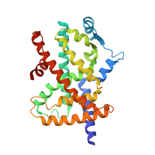
    Covalent modification of proteins is important for normal cellular regulation. Here, we report on the covalent modification of peroxisome proliferator-activated receptor γ (PPARγ), an important drug target, by oxo-fatty acids. In this study, ESI mass spectroscopy showed that the reactivities of oxo-fatty acids with PPARγ are different from one another and that these behaviors are related to the structure of the fatty acids. X-ray crystallography showed that three oxo-fatty acids all bound to the same residue of PPARγ (Cys285), but displayed different hydrogen bonding modes. Moreover, fatty acids formed covalent bonds with both PPARγ moieties in the homodimer, one in an active conformation and the other in an alternative conformation. These two conformations may explain why covalently bound fatty acids show partial rather than full agonist activity.


  • Organizational Affiliation

    Laboratory of Drug Design and Medicinal Chemistry, Showa Pharmaceutical University, 3-3165 Higashi-Tamagawagakuen, Machida, Tokyo 194-8543, Japan.


Macromolecules
Find similar proteins by:  (by identity cutoff)  |  3D Structure
Entity ID: 1
MoleculeChains Sequence LengthOrganismDetailsImage
Peroxisome proliferator-activated receptor gamma
A, B
276Homo sapiensMutation(s): 0 
Gene Names: PPARGNR1C3
UniProt & NIH Common Fund Data Resources
Find proteins for P37231 (Homo sapiens)
Explore P37231 
Go to UniProtKB:  P37231
PHAROS:  P37231
GTEx:  ENSG00000132170 
Entity Groups  
Sequence Clusters30% Identity50% Identity70% Identity90% Identity95% Identity100% Identity
UniProt GroupP37231
Sequence Annotations
Expand
  • Reference Sequence
Small Molecules
Ligands 1 Unique
IDChains Name / Formula / InChI Key2D Diagram3D Interactions
66B
Query on 66B

Download Ideal Coordinates CCD File 
C [auth A],
D [auth B]
(8E,12Z,15Z,18Z,21Z)-6-oxotetracosa-8,12,15,18,21-pentaenoic acid
C24 H36 O3
SOUDNDRWZSIFBW-IIFHDYRKSA-N
Experimental Data & Validation

Experimental Data

  • Method: X-RAY DIFFRACTION
  • Resolution: 2.40 Å
  • R-Value Free: 0.279 
  • R-Value Work: 0.229 
  • R-Value Observed: 0.231 
  • Space Group: C 1 2 1
Unit Cell:
Length ( Å )Angle ( ˚ )
a = 93.34α = 90
b = 61.92β = 102.72
c = 118.82γ = 90
Software Package:
Software NamePurpose
SERGUIdata collection
REFMACrefinement
MOSFLMdata reduction
SCALAdata scaling

Structure Validation

View Full Validation Report


Ligand Structure Quality Assessment 


Entry History 

Deposition Data

Revision History  (Full details and data files)

  • Version 1.0: 2015-04-08
    Type: Initial release
  • Version 1.1: 2017-11-22
    Changes: Refinement description
  • Version 1.2: 2022-08-24
    Changes: Database references, Derived calculations