3WZE

KDR in complex with ligand sorafenib


Experimental Data Snapshot

  • Method: X-RAY DIFFRACTION
  • Resolution: 1.90 Å
  • R-Value Free: 0.224 
  • R-Value Work: 0.185 
  • R-Value Observed: 0.188 

wwPDB Validation   3D Report Full Report


Ligand Structure Quality Assessment 


This is version 1.0 of the entry. See complete history


Literature

Distinct binding mode of multikinase inhibitor lenvatinib revealed by biochemical characterization.

Okamoto, K.Ikemori-Kawada, M.Jestel, A.von Konig, K.Funahashi, Y.Matsushima, T.Tsuruoka, A.Inoue, A.Matsui, J.

(2015) ACS Med Chem Lett 6: 89-94

  • DOI: https://doi.org/10.1021/ml500394m
  • Primary Citation of Related Structures:  
    3WZD, 3WZE

  • PubMed Abstract: 

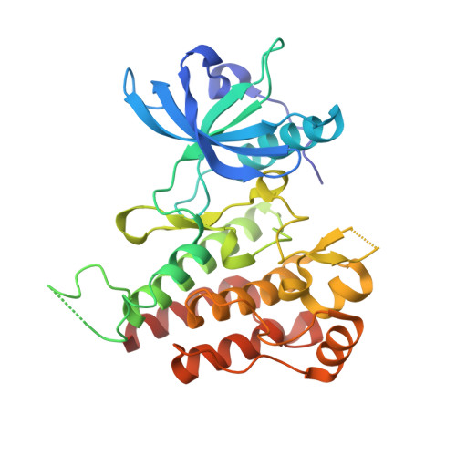
    Lenvatinib is an oral multikinase inhibitor that selectively inhibits vascular endothelial growth factor (VEGF) receptors 1 to 3 and other proangiogenic and oncogenic pathway-related receptor tyrosine kinases. To elucidate the origin of the potency of lenvatinib in VEGF receptor 2 (VEGFR2) inhibition, we conducted a kinetic interaction analysis of lenvatinib with VEGFR2 and X-ray analysis of the crystal structure of VEGFR2-lenvatinib complexes. Kinetic analysis revealed that lenvatinib had a rapid association rate constant and a relatively slow dissociation rate constant in complex with VEGFR2. Co-crystal structure analysis demonstrated that lenvatinib binds at its ATP mimetic quinoline moiety to the ATP binding site and to the neighboring region via a cyclopropane ring, adopting an Asp-Phe-Gly (DFG)-"in" conformation. These results suggest that lenvatinib is very distinct in its binding mode of interaction compared to the several approved VEGFR2 kinase inhibitors.


  • Organizational Affiliation

    Eisai Co., Ltd , Tsukuba, Ibaraki 300-2635, Japan.


Macromolecules
Find similar proteins by:  (by identity cutoff)  |  3D Structure
Entity ID: 1
MoleculeChains Sequence LengthOrganismDetailsImage
Vascular endothelial growth factor receptor 2309Homo sapiensMutation(s): 1 
Gene Names: KDRFLK1VEGFR2
EC: 2.7.10.1
UniProt & NIH Common Fund Data Resources
Find proteins for P35968 (Homo sapiens)
Explore P35968 
Go to UniProtKB:  P35968
PHAROS:  P35968
GTEx:  ENSG00000128052 
Entity Groups  
Sequence Clusters30% Identity50% Identity70% Identity90% Identity95% Identity100% Identity
UniProt GroupP35968
Sequence Annotations
Expand
  • Reference Sequence
Small Molecules
Ligands 3 Unique
IDChains Name / Formula / InChI Key2D Diagram3D Interactions
BAX
Query on BAX

Download Ideal Coordinates CCD File 
B [auth A]4-{4-[({[4-CHLORO-3-(TRIFLUOROMETHYL)PHENYL]AMINO}CARBONYL)AMINO]PHENOXY}-N-METHYLPYRIDINE-2-CARBOXAMIDE
C21 H16 Cl F3 N4 O3
MLDQJTXFUGDVEO-UHFFFAOYSA-N
DTT
Query on DTT

Download Ideal Coordinates CCD File 
D [auth A]2,3-DIHYDROXY-1,4-DITHIOBUTANE
C4 H10 O2 S2
VHJLVAABSRFDPM-IMJSIDKUSA-N
ACT
Query on ACT

Download Ideal Coordinates CCD File 
C [auth A]ACETATE ION
C2 H3 O2
QTBSBXVTEAMEQO-UHFFFAOYSA-M
Binding Affinity Annotations 
IDSourceBinding Affinity
BAX BindingDB:  3WZE Ki: min: 0.02, max: 22 (nM) from 3 assay(s)
Kd: min: 33, max: 93 (nM) from 3 assay(s)
IC50: min: 0.16, max: 1210 (nM) from 44 assay(s)
EC50: 500 (nM) from 1 assay(s)
Binding MOAD:  3WZE Kd: 33 (nM) from 1 assay(s)
Experimental Data & Validation

Experimental Data

  • Method: X-RAY DIFFRACTION
  • Resolution: 1.90 Å
  • R-Value Free: 0.224 
  • R-Value Work: 0.185 
  • R-Value Observed: 0.188 
  • Space Group: C 1 2 1
Unit Cell:
Length ( Å )Angle ( ˚ )
a = 135.921α = 90
b = 57.357β = 94.17
c = 52.17γ = 90
Software Package:
Software NamePurpose
REFMACrefinement
XDSdata reduction
XSCALEdata scaling

Structure Validation

View Full Validation Report


Ligand Structure Quality Assessment 


Entry History 

Deposition Data

Revision History  (Full details and data files)

  • Version 1.0: 2015-05-27
    Type: Initial release