


Structure of calcium bound cutinase Est119 from Thermobifida alba.
Kitadokoro, K., Thumarat, U., Kawai, F.To be published.
Experimental Data Snapshot
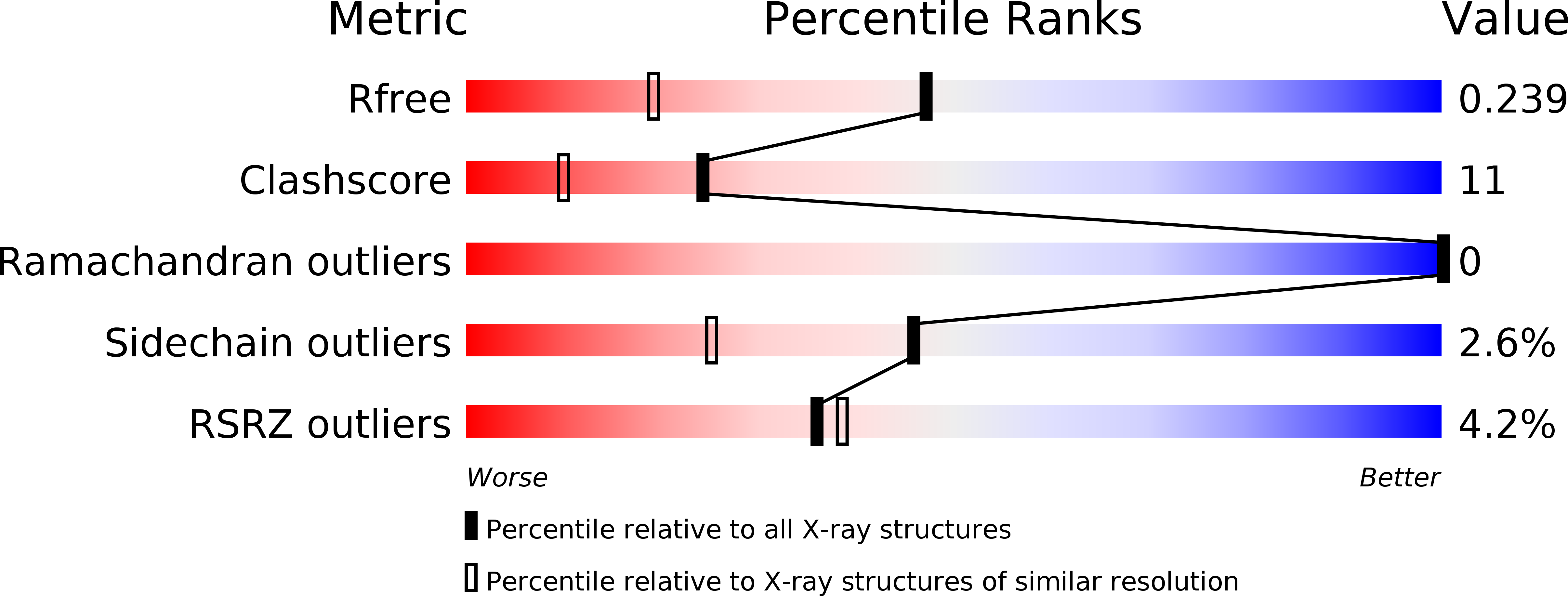
wwPDB Validation 3D Report Full Report
Entity ID: 1 | |||||
|---|---|---|---|---|---|
| Molecule | Chains | Sequence Length | Organism | Details | Image |
| Esterase | 306 | Thermobifida alba | Mutation(s): 0 Gene Names: est2 | 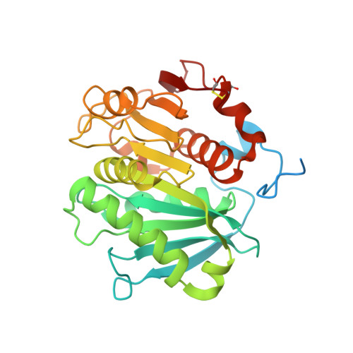 | |
UniProt | |||||
Find proteins for F7IX06 (Thermobifida alba) Explore F7IX06 Go to UniProtKB: F7IX06 | |||||
Entity Groups | |||||
| Sequence Clusters | 30% Identity50% Identity70% Identity90% Identity95% Identity100% Identity | ||||
| UniProt Group | F7IX06 | ||||
Sequence AnnotationsExpand | |||||
| |||||
| Ligands 2 Unique | |||||
|---|---|---|---|---|---|
| ID | Chains | Name / Formula / InChI Key | 2D Diagram | 3D Interactions | |
| 2PE Query on 2PE | D [auth B] | NONAETHYLENE GLYCOL C18 H38 O10 YZUUTMGDONTGTN-UHFFFAOYSA-N |  | ||
| CA Query on CA | C [auth A], E [auth B], F [auth B] | CALCIUM ION Ca BHPQYMZQTOCNFJ-UHFFFAOYSA-N |  | ||
| Length ( Å ) | Angle ( ˚ ) |
|---|---|
| a = 101.473 | α = 90 |
| b = 87.74 | β = 133.35 |
| c = 72.619 | γ = 90 |
| Software Name | Purpose |
|---|---|
| HKL-2000 | data collection |
| MOLREP | phasing |
| REFMAC | refinement |
| HKL-2000 | data reduction |
| HKL-2000 | data scaling |
RCSB PDB (citation) is hosted by
RCSB PDB is a member of the

