3WWR

Crystal structure of the human vitamin D receptor ligand binding domain complexed with 1-((((1S,2R,6R,Z)-2,6-dihydroxy-4-((E)-2-((1R,3aS,7aR)-1-((R)-6-hydroxy-6-methylheptan-2-yl)-7a-methylhexahydro-1H-inden-4(2H)-ylidene)ethylidene)-3-methylenecyclohexyl)oxy)methyl)cyclopropanecarbonitrile


Experimental Data Snapshot

  • Method: X-RAY DIFFRACTION
  • Resolution: 3.18 Å
  • R-Value Free: 0.291 
  • R-Value Work: 0.247 
  • R-Value Observed: 0.249 

wwPDB Validation   3D Report Full Report


Ligand Structure Quality Assessment 


This is version 1.3 of the entry. See complete history


Literature

Synthesis and biological activities of vitamin D3 derivatives with cyanoalkyl side chain at C-2 position.

Saitoh, H.Watanabe, H.Kakuda, S.Takimoto-Kamimura, M.Takagi, K.Takeuchi, A.Takenouchi, K.

(2015) J Steroid Biochem Mol Biol 148: 27-30

  • DOI: https://doi.org/10.1016/j.jsbmb.2014.12.004
  • Primary Citation of Related Structures:  
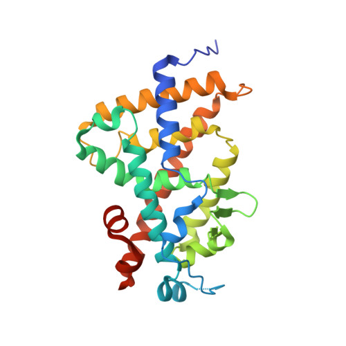
    3WWR

  • PubMed Abstract: 

    We synthesized and evaluated novel vitamin D3 derivatives with cyanoalkyl side chain at C-2 position on the basis of our previous research for 2α side chain which bears nitrogen atom-containing functional group. Through a study of X-ray co-crystal structures of human VDR and compound 3, we demonstrated that the 2α alkyl side chain in compound 3 shows a novel interaction in the complex of hVDR-LBD and ligand. This article is part of a Special Issue entitled '17th Vitamin D Workshop'.


  • Organizational Affiliation

    Teijin Institute for Bio-Medical Research, Teijin Pharma Ltd., 4-3-2 Asahigaoka, Hino, Tokyo 191-8512, Japan. Electronic address: hi.saitou@teijin.co.jp.


Macromolecules
Find similar proteins by:  (by identity cutoff)  |  3D Structure
Entity ID: 1
MoleculeChains Sequence LengthOrganismDetailsImage
Vitamin D3 receptor263Homo sapiensMutation(s): 0 
Gene Names: VDRNR1I1
UniProt & NIH Common Fund Data Resources
Find proteins for P11473 (Homo sapiens)
Explore P11473 
Go to UniProtKB:  P11473
PHAROS:  P11473
GTEx:  ENSG00000111424 
Entity Groups  
Sequence Clusters30% Identity50% Identity70% Identity90% Identity95% Identity100% Identity
UniProt GroupP11473
Sequence Annotations
Expand
  • Reference Sequence
Small Molecules
Ligands 1 Unique
IDChains Name / Formula / InChI Key2D Diagram3D Interactions
3AJ
Query on 3AJ

Download Ideal Coordinates CCD File 
B [auth A]1-({[(1R,2S,3R,5Z,7E,14beta,17alpha)-1,3,25-trihydroxy-9,10-secocholesta-5,7,10-trien-2-yl]oxy}methyl)cyclopropanecarbonitrile
C32 H49 N O4
XRVHRGHHDVJYGR-IPBGMLIISA-N
Experimental Data & Validation

Experimental Data

  • Method: X-RAY DIFFRACTION
  • Resolution: 3.18 Å
  • R-Value Free: 0.291 
  • R-Value Work: 0.247 
  • R-Value Observed: 0.249 
  • Space Group: P 21 21 21
Unit Cell:
Length ( Å )Angle ( ˚ )
a = 45.167α = 90
b = 51.826β = 90
c = 132.384γ = 90
Software Package:
Software NamePurpose
HKL-2000data collection
MOLREPphasing
REFMACrefinement
HKL-2000data reduction
HKL-2000data scaling

Structure Validation

View Full Validation Report


Ligand Structure Quality Assessment 


Entry History 

Deposition Data

Revision History  (Full details and data files)

  • Version 1.0: 2014-12-31
    Type: Initial release
  • Version 1.1: 2017-08-16
    Changes: Refinement description, Source and taxonomy
  • Version 1.2: 2022-08-24
    Changes: Database references, Derived calculations
  • Version 1.3: 2023-11-08
    Changes: Data collection, Refinement description