3WQH

Crystal Structure of human DPP-IV in complex with Anagliptin


Experimental Data Snapshot

  • Method: X-RAY DIFFRACTION
  • Resolution: 2.85 Å
  • R-Value Free: 0.288 
  • R-Value Work: 0.227 

wwPDB Validation   3D Report Full Report


Ligand Structure Quality Assessment 


This is version 1.3 of the entry. See complete history


Literature

Anagliptin, a potent dipeptidyl peptidase IV inhibitor: its single-crystal structure and enzyme interactions.

Watanabe, Y.S.Yasuda, Y.Kojima, Y.Okada, S.Motoyama, T.Takahashi, R.Oka, M.

(2015) J Enzyme Inhib Med Chem 30: 981-988

  • DOI: https://doi.org/10.3109/14756366.2014.1002402
  • Primary Citation of Related Structures:  
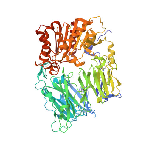
    3WQH

  • PubMed Abstract: 

    The single-crystal structure of anagliptin, N-[2-({2-[(2S)-2-cyanopyrrolidin-1-yl]-2-oxoethyl}amino)-2-methylpropyl]-2-methylpyrazolo[1,5-a]pyrimidine-6-carboxamide, was determined. Two independent molecules were held together by intermolecular hydrogen bonds, and the absolute configuration of the 2-cyanopyrrolidine ring delivered from l-prolinamide was confirmed to be S. The interactions of anagliptin with DPP-4 were clarified by the co-crystal structure solved at 2.85 Å resolution. Based on the structure determined by X-ray crystallography, the potency and selectivity of anagliptin were discussed, and an SAR study using anagliptin derivatives was performed.


  • Organizational Affiliation

    a Mie Research Park, Sanwa Kagaku Kenkyusho Co., Ltd. , Mie , Japan .


Macromolecules
Find similar proteins by:  (by identity cutoff)  |  3D Structure
Entity ID: 1
MoleculeChains Sequence LengthOrganismDetailsImage
Dipeptidyl peptidase 4
A, B
730Homo sapiensMutation(s): 0 
Gene Names: DPP4ADCP2CD26
EC: 3.4.14.5
UniProt & NIH Common Fund Data Resources
Find proteins for P27487 (Homo sapiens)
Explore P27487 
Go to UniProtKB:  P27487
PHAROS:  P27487
GTEx:  ENSG00000197635 
Entity Groups  
Sequence Clusters30% Identity50% Identity70% Identity90% Identity95% Identity100% Identity
UniProt GroupP27487
Sequence Annotations
Expand
  • Reference Sequence
Small Molecules
Ligands 2 Unique
IDChains Name / Formula / InChI Key2D Diagram3D Interactions
SKK
Query on SKK

Download Ideal Coordinates CCD File 
J [auth A],
Q [auth B]
N-[2-({2-[(2S)-2-cyanopyrrolidin-1-yl]-2-oxoethyl}amino)-2-methylpropyl]-2-methylpyrazolo[1,5-a]pyrimidine-6-carboxamide
C19 H25 N7 O2
LDXYBEHACFJIEL-HNNXBMFYSA-N
NAG
Query on NAG

Download Ideal Coordinates CCD File 
C [auth A]
D [auth A]
E [auth A]
F [auth A]
G [auth A]
C [auth A],
D [auth A],
E [auth A],
F [auth A],
G [auth A],
H [auth A],
I [auth A],
K [auth B],
L [auth B],
M [auth B],
N [auth B],
O [auth B],
P [auth B]
2-acetamido-2-deoxy-beta-D-glucopyranose
C8 H15 N O6
OVRNDRQMDRJTHS-FMDGEEDCSA-N
Binding Affinity Annotations 
IDSourceBinding Affinity
SKK BindingDB:  3WQH IC50: 3.8 (nM) from 1 assay(s)
Experimental Data & Validation

Experimental Data

  • Method: X-RAY DIFFRACTION
  • Resolution: 2.85 Å
  • R-Value Free: 0.288 
  • R-Value Work: 0.227 
  • Space Group: P 21 21 21
Unit Cell:
Length ( Å )Angle ( ˚ )
a = 65.16α = 90
b = 68.99β = 90
c = 419.6γ = 90
Software Package:
Software NamePurpose
XDSdata scaling
MOLREPphasing
PRIME-Xrefinement
XDSdata reduction
XSCALEdata scaling

Structure Validation

View Full Validation Report


Ligand Structure Quality Assessment 


Entry History 

Deposition Data

Revision History  (Full details and data files)

  • Version 1.0: 2015-07-15
    Type: Initial release
  • Version 1.1: 2020-07-29
    Type: Remediation
    Reason: Carbohydrate remediation
    Changes: Data collection, Database references, Derived calculations, Structure summary
  • Version 1.2: 2022-08-24
    Changes: Database references, Refinement description, Structure summary
  • Version 1.3: 2023-11-08
    Changes: Data collection, Refinement description