3WKY

Crystal structure of hemolymph type prophenoloxidase (proPOb) from crustacean


Experimental Data Snapshot

  • Method: X-RAY DIFFRACTION
  • Resolution: 1.80 Å
  • R-Value Free: 0.196 
  • R-Value Work: 0.175 
  • R-Value Observed: 0.176 

wwPDB Validation   3D Report Full Report


Ligand Structure Quality Assessment 


This is version 2.1 of the entry. See complete history


Literature

The crystal structure of a crustacean prophenoloxidase provides a clue to understanding the functionality of the type 3 copper proteins.

Masuda, T.Momoji, K.Hirata, T.Mikami, B.

(2014) FEBS J 281: 2659-2673

  • DOI: https://doi.org/10.1111/febs.12812
  • Primary Citation of Related Structures:  
    3WKY

  • PubMed Abstract: 

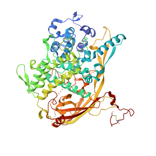
    Phenoloxidase (PO), which is classified as a type 3 copper protein, catalyzes the hydroxylation of monophenol to o-diphenol and subsequent oxidation to the corresponding o-quinone. The geometry and coordination environment of the active site of the arthropod PO are very similar to those of the arthropod hemocyanin (Hc). However, unlike the POs, Hc is an oxygen carrier in crustaceans, and does not possess PO activity in general. Recently, we identified a new type of proPO from a crustacean and designated it proPOβ. This enzyme has many characteristics that are rather similar to those of Hc, such as its maturation, localization, and oligomeric state. Here, we determined the crystal structure of proPOβ prepared from the hemolymph of kuruma prawns (Marsupenaeus japonicus) at 1.8-Å resolution. M. japonicus proPOβ forms a homohexamer rather similar to that of arthropod Hc. The geometry of the active copper site in proPOβ is nearly identical to that of arthropod Hc. Furthermore, the well-characterized 'place-holder' phenylalanine is present (Phe72). However, the accessibility to the active site differs in several ways. First, another phenylalanine, which shields the active site by interacting with a copper-coordinated histidine in crustacean Hc, is replaced by valine in the proPOβ structure. Second, two tyrosines, Tyr208 and Tyr209, both of which are absent in Hc, show the alternative conformations and form a pathway providing access to the reaction center. Thus, the present crystal structure clarifies the similarities and differences in the activity of two closely related proteins, PO and Hc.


  • Organizational Affiliation

    Laboratory of Food Quality Design and Development, Division of Agronomy and Horticultural Science, Graduate School of Agriculture, Kyoto University, Japan.


Macromolecules
Find similar proteins by:  (by identity cutoff)  |  3D Structure
Entity ID: 1
MoleculeChains Sequence LengthOrganismDetailsImage
Prophenoloxidase b
A, B
687Penaeus japonicusMutation(s): 0 
UniProt
Find proteins for G5EKM4 (Penaeus japonicus)
Explore G5EKM4 
Go to UniProtKB:  G5EKM4
Entity Groups  
Sequence Clusters30% Identity50% Identity70% Identity90% Identity95% Identity100% Identity
UniProt GroupG5EKM4
Sequence Annotations
Expand
  • Reference Sequence
Oligosaccharides

Help

Entity ID: 2
MoleculeChains Length2D Diagram Glycosylation3D Interactions
2-acetamido-2-deoxy-beta-D-glucopyranose-(1-4)-2-acetamido-2-deoxy-beta-D-glucopyranose
C, D
2N-Glycosylation
Glycosylation Resources
GlyTouCan:  G42666HT
GlyCosmos:  G42666HT
GlyGen:  G42666HT
Small Molecules
Ligands 6 Unique
IDChains Name / Formula / InChI Key2D Diagram3D Interactions
NAG
Query on NAG

Download Ideal Coordinates CCD File 
E [auth A]
F [auth A]
G [auth A]
H [auth A]
I [auth A]
E [auth A],
F [auth A],
G [auth A],
H [auth A],
I [auth A],
U [auth B],
V [auth B],
W [auth B],
X [auth B],
Y [auth B]
2-acetamido-2-deoxy-beta-D-glucopyranose
C8 H15 N O6
OVRNDRQMDRJTHS-FMDGEEDCSA-N
CUO
Query on CUO

Download Ideal Coordinates CCD File 
J [auth A],
Z [auth B]
CU2-O2 CLUSTER
Cu2 O2
PCGDDKBKPXANNY-UHFFFAOYSA-N
EDO
Query on EDO

Download Ideal Coordinates CCD File 
AA [auth B]
BA [auth B]
CA [auth B]
DA [auth B]
K [auth A]
AA [auth B],
BA [auth B],
CA [auth B],
DA [auth B],
K [auth A],
L [auth A],
M [auth A],
N [auth A],
O [auth A]
1,2-ETHANEDIOL
C2 H6 O2
LYCAIKOWRPUZTN-UHFFFAOYSA-N
CL
Query on CL

Download Ideal Coordinates CCD File 
FA [auth B],
Q [auth A],
R [auth A],
S [auth A],
T [auth A]
CHLORIDE ION
Cl
VEXZGXHMUGYJMC-UHFFFAOYSA-M
MG
Query on MG

Download Ideal Coordinates CCD File 
EA [auth B]MAGNESIUM ION
Mg
JLVVSXFLKOJNIY-UHFFFAOYSA-N
NA
Query on NA

Download Ideal Coordinates CCD File 
P [auth A]SODIUM ION
Na
FKNQFGJONOIPTF-UHFFFAOYSA-N
Experimental Data & Validation

Experimental Data

  • Method: X-RAY DIFFRACTION
  • Resolution: 1.80 Å
  • R-Value Free: 0.196 
  • R-Value Work: 0.175 
  • R-Value Observed: 0.176 
  • Space Group: H 3
Unit Cell:
Length ( Å )Angle ( ˚ )
a = 156.706α = 90
b = 156.706β = 90
c = 283.83γ = 120
Software Package:
Software NamePurpose
HKL-2000data collection
SOLVEphasing
PHENIXrefinement
HKL-2000data reduction
HKL-2000data scaling

Structure Validation

View Full Validation Report


Ligand Structure Quality Assessment 


Entry History 

Deposition Data

Revision History  (Full details and data files)

  • Version 1.0: 2014-04-23
    Type: Initial release
  • Version 2.0: 2020-07-29
    Type: Remediation
    Reason: Carbohydrate remediation
    Changes: Atomic model, Data collection, Derived calculations, Structure summary
  • Version 2.1: 2022-08-24
    Changes: Database references, Structure summary