3WKT

Complex structure of an open form of NADPH-cytochrome P450 reductase and heme oxygenase-1


Experimental Data Snapshot

  • Method: X-RAY DIFFRACTION
  • Resolution: 4.30 Å
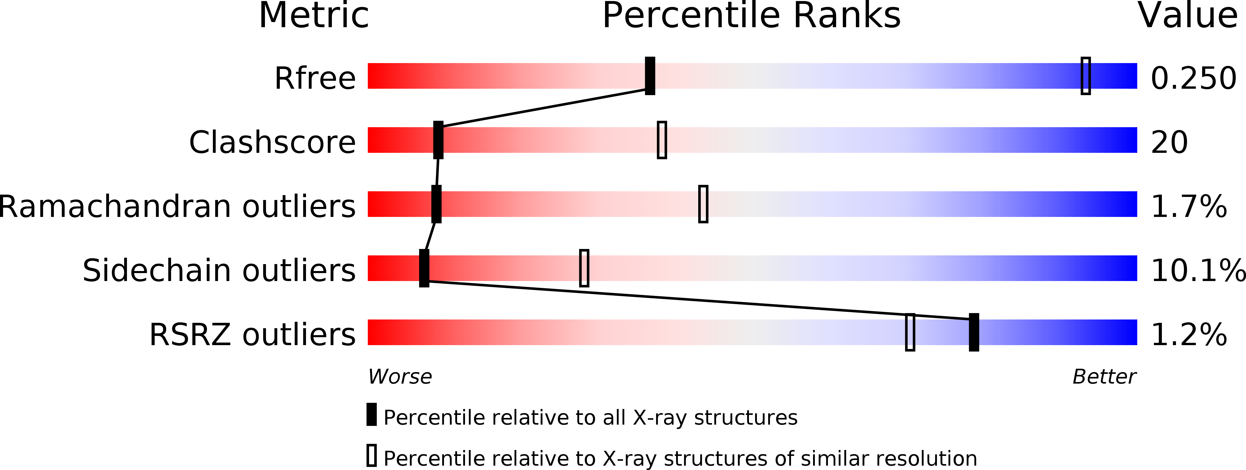
  • R-Value Free: 0.256 
  • R-Value Work: 0.222 
  • R-Value Observed: 0.224 

wwPDB Validation   3D Report Full Report


Ligand Structure Quality Assessment 


This is version 1.2 of the entry. See complete history


Literature

Structural basis for the electron transfer from an open form of NADPH-cytochrome P450 oxidoreductase to heme oxygenase.

Sugishima, M.Sato, H.Higashimoto, Y.Harada, J.Wada, K.Fukuyama, K.Noguchi, M.

(2014) Proc Natl Acad Sci U S A 111: 2524-2529

  • DOI: https://doi.org/10.1073/pnas.1322034111
  • Primary Citation of Related Structures:  
    3WKT

  • PubMed Abstract: 

    NADPH-cytochrome P450 oxidoreductase (CPR) supplies electrons to various heme proteins including heme oxygenase (HO), which is a key enzyme for heme degradation. Electrons from NADPH flow first to flavin adenine dinucleotide, then to flavin mononucleotide (FMN), and finally to heme in the redox partner. For electron transfer from CPR to its redox partner, the ''closed-open transition'' of CPR is indispensable. Here, we demonstrate that a hinge-shortened CPR variant, which favors an open conformation, makes a stable complex with heme-HO-1 and can support the HO reaction, although its efficiency is extremely limited. Furthermore, we determined the crystal structure of the CPR variant in complex with heme-HO-1 at 4.3-Å resolution. The crystal structure of a complex of CPR and its redox partner was previously unidentified. The distance between heme and FMN in this complex (6 Å) implies direct electron transfer from FMN to heme.


  • Organizational Affiliation

    Department of Medical Biochemistry and Department of Chemistry, Kurume University School of Medicine, Kurume, Fukuoka 830-0011, Japan.


Macromolecules
Find similar proteins by:  (by identity cutoff)  |  3D Structure
Entity ID: 1
MoleculeChains Sequence LengthOrganismDetailsImage
NADPH-cytochrome P450 reductase
A, B
618Rattus norvegicusMutation(s): 0 
Gene Names: Por
EC: 1.6.2.4
UniProt
Find proteins for P00388 (Rattus norvegicus)
Explore P00388 
Go to UniProtKB:  P00388
Entity Groups  
Sequence Clusters30% Identity50% Identity70% Identity90% Identity95% Identity100% Identity
UniProt GroupP00388
Sequence Annotations
Expand
  • Reference Sequence
Find similar proteins by:  (by identity cutoff)  |  3D Structure
Entity ID: 2
MoleculeChains Sequence LengthOrganismDetailsImage
Heme oxygenase 1
C, D
267Rattus norvegicusMutation(s): 0 
Gene Names: Hmox1
EC: 1.14.99.3
UniProt
Find proteins for P06762 (Rattus norvegicus)
Explore P06762 
Go to UniProtKB:  P06762
Entity Groups  
Sequence Clusters30% Identity50% Identity70% Identity90% Identity95% Identity100% Identity
UniProt GroupP06762
Sequence Annotations
Expand
  • Reference Sequence
Small Molecules
Ligands 4 Unique
IDChains Name / Formula / InChI Key2D Diagram3D Interactions
FAD
Query on FAD

Download Ideal Coordinates CCD File 
E [auth A],
H [auth B]
FLAVIN-ADENINE DINUCLEOTIDE
C27 H33 N9 O15 P2
VWWQXMAJTJZDQX-UYBVJOGSSA-N
NAP
Query on NAP

Download Ideal Coordinates CCD File 
G [auth A],
J [auth B]
NADP NICOTINAMIDE-ADENINE-DINUCLEOTIDE PHOSPHATE
C21 H28 N7 O17 P3
XJLXINKUBYWONI-NNYOXOHSSA-N
HEM
Query on HEM

Download Ideal Coordinates CCD File 
K [auth C],
L [auth D]
PROTOPORPHYRIN IX CONTAINING FE
C34 H32 Fe N4 O4
KABFMIBPWCXCRK-RGGAHWMASA-L
FMN
Query on FMN

Download Ideal Coordinates CCD File 
F [auth A],
I [auth B]
FLAVIN MONONUCLEOTIDE
C17 H21 N4 O9 P
FVTCRASFADXXNN-SCRDCRAPSA-N
Experimental Data & Validation

Experimental Data

  • Method: X-RAY DIFFRACTION
  • Resolution: 4.30 Å
  • R-Value Free: 0.256 
  • R-Value Work: 0.222 
  • R-Value Observed: 0.224 
  • Space Group: P 61
Unit Cell:
Length ( Å )Angle ( ˚ )
a = 290.336α = 90
b = 290.336β = 90
c = 83.646γ = 120
Software Package:
Software NamePurpose
BSSdata collection
MOLREPphasing
REFMACrefinement
HKL-2000data reduction
HKL-2000data scaling

Structure Validation

View Full Validation Report


Ligand Structure Quality Assessment 


Entry History 

Deposition Data

Revision History  (Full details and data files)

  • Version 1.0: 2014-01-29
    Type: Initial release
  • Version 1.1: 2016-06-01
    Changes: Database references
  • Version 1.2: 2023-11-08
    Changes: Data collection, Database references, Derived calculations, Refinement description