


3WIR
Crystal structure of kojibiose phosphorylase complexed with glucose
- PDB DOI: https://doi.org/10.2210/pdb3WIR/pdb
- Classification: TRANSFERASE
- Organism(s): Caldicellulosiruptor saccharolyticus DSM 8903
- Expression System: Escherichia coli
- Mutation(s): No
- Deposited: 2013-09-24 Released: 2014-02-05
Experimental Data Snapshot
- Method: X-RAY DIFFRACTION
- Resolution: 2.05 Å
- R-Value Free: 0.251
- R-Value Work: 0.191
- R-Value Observed: 0.194

This is version 1.2 of the entry. See complete history.

Macromolecules
Find similar proteins by:
(by identity cutoff) | 3D Structure
Entity ID: 1 | |||||
|---|---|---|---|---|---|
| Molecule | Chains | Sequence Length | Organism | Details | Image |
| Kojibiose phosphorylase | 764 | Caldicellulosiruptor saccharolyticus DSM 8903 | Mutation(s): 0 Gene Names: Csac_0444 EC: 2.4.1.230 | 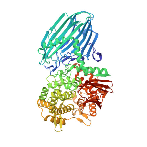 | |
UniProt | |||||
Find proteins for A4XGP2 (Caldicellulosiruptor saccharolyticus (strain ATCC 43494 / DSM 8903 / Tp8T 6331)) Explore A4XGP2 Go to UniProtKB: A4XGP2 | |||||
Entity Groups | |||||
| Sequence Clusters | 30% Identity50% Identity70% Identity90% Identity95% Identity100% Identity | ||||
| UniProt Group | A4XGP2 | ||||
Sequence AnnotationsExpand | |||||
| |||||
Small Molecules
| Ligands 3 Unique | |||||
|---|---|---|---|---|---|
| ID | Chains | Name / Formula / InChI Key | 2D Diagram | 3D Interactions | |
| BGC Query on BGC | E [auth A] F [auth A] J [auth B] K [auth B] O [auth C] | beta-D-glucopyranose C6 H12 O6 WQZGKKKJIJFFOK-VFUOTHLCSA-N |  | ||
| PO4 Query on PO4 | AA [auth D] I [auth A] M [auth B] N [auth B] S [auth C] | PHOSPHATE ION O4 P NBIIXXVUZAFLBC-UHFFFAOYSA-K |  | ||
| GOL Query on GOL | G [auth A] H [auth A] L [auth B] Q [auth C] R [auth C] | GLYCEROL C3 H8 O3 PEDCQBHIVMGVHV-UHFFFAOYSA-N |  | ||
Experimental Data & Validation
Experimental Data
- Method: X-RAY DIFFRACTION
- Resolution: 2.05 Å
- R-Value Free: 0.251
- R-Value Work: 0.191
- R-Value Observed: 0.194
- Space Group: P 1
Unit Cell:
| Length ( Å ) | Angle ( ˚ ) |
|---|---|
| a = 71.581 | α = 68.83 |
| b = 104.464 | β = 86.02 |
| c = 124.164 | γ = 90.06 |
| Software Name | Purpose |
|---|---|
| HKL-2000 | data collection |
| MOLREP | phasing |
| REFMAC | refinement |
| HKL-2000 | data reduction |
| HKL-2000 | data scaling |
Entry History
Deposition Data
- Released Date: 2014-02-05 Deposition Author(s): Okada, S., Yamamoto, T., Watanabe, H., Nishimoto, T., Chaen, H., Fukuda, S., Wakagi, T., Fushinobu, S.
Revision History (Full details and data files)
- Version 1.0: 2014-02-05
Type: Initial release - Version 1.1: 2020-07-29
Type: Remediation
Reason: Carbohydrate remediation
Changes: Data collection, Database references, Derived calculations, Structure summary - Version 1.2: 2023-11-08
Changes: Data collection, Database references, Refinement description, Structure summary
