3WI3

Crystal Structure of the Sld3/Treslin domain from yeast Sld3


Experimental Data Snapshot

  • Method: X-RAY DIFFRACTION
  • Resolution: 2.40 Å
  • R-Value Free: 0.262 
  • R-Value Work: 0.217 
  • R-Value Observed: 0.219 

wwPDB Validation   3D Report Full Report


This is version 1.2 of the entry. See complete history


Literature

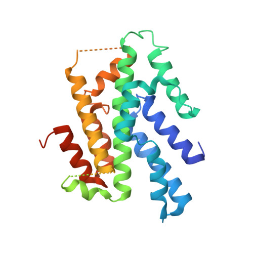
Crystal structure of the homology domain of the eukaryotic DNA replication proteins sld3/treslin.

Itou, H.Muramatsu, S.Shirakihara, Y.Araki, H.

(2014) Structure 22: 1341-1347

  • DOI: https://doi.org/10.1016/j.str.2014.07.001
  • Primary Citation of Related Structures:  
    3WI3

  • PubMed Abstract: 

    The initiation of eukaryotic chromosomal DNA replication requires the formation of an active replicative helicase at the replication origins of chromosomal DNA. Yeast Sld3 and its metazoan counterpart Treslin are the hub proteins mediating protein associations critical for the helicase formation. Here, we show the crystal structure of the central domain of Sld3 that is conserved in Sld3/Treslin family of proteins. The domain consists of two segments with 12 helices and is sufficient to bind to Cdc45, the essential helicase component. The structure model of the Sld3-Cdc45 complex, which is crucial for the formation of the active helicase, is proposed.


  • Organizational Affiliation

    Structural Biology Center, National Institute of Genetics, Yata1111, Mishima, Shizuoka 411-8540, Japan; Department of Genetics, SOKENDAI, Yata1111, Mishima, Shizuoka 411-8540, Japan. Electronic address: hitou@nig.ac.jp.


Macromolecules
Find similar proteins by:  (by identity cutoff)  |  3D Structure
Entity ID: 1
MoleculeChains Sequence LengthOrganismDetailsImage
DNA replication regulator SLD3
A, B, C
292Saccharomyces cerevisiae S288CMutation(s): 0 
Gene Names: SLD3YGL113WG2980
UniProt
Find proteins for P53135 (Saccharomyces cerevisiae (strain ATCC 204508 / S288c))
Explore P53135 
Go to UniProtKB:  P53135
Entity Groups  
Sequence Clusters30% Identity50% Identity70% Identity90% Identity95% Identity100% Identity
UniProt GroupP53135
Sequence Annotations
Expand
  • Reference Sequence
Small Molecules
Ligands 2 Unique
IDChains Name / Formula / InChI Key2D Diagram3D Interactions
SO4
Query on SO4

Download Ideal Coordinates CCD File 
D [auth A],
DA [auth C],
U [auth B]
SULFATE ION
O4 S
QAOWNCQODCNURD-UHFFFAOYSA-L
EDO
Query on EDO

Download Ideal Coordinates CCD File 
AA [auth B]
BA [auth B]
CA [auth B]
E [auth A]
F [auth A]
AA [auth B],
BA [auth B],
CA [auth B],
E [auth A],
F [auth A],
G [auth A],
H [auth A],
I [auth A],
J [auth A],
K [auth A],
L [auth A],
M [auth A],
N [auth A],
O [auth A],
P [auth A],
Q [auth A],
R [auth A],
S [auth A],
T [auth A],
V [auth B],
W [auth B],
X [auth B],
Y [auth B],
Z [auth B]
1,2-ETHANEDIOL
C2 H6 O2
LYCAIKOWRPUZTN-UHFFFAOYSA-N
Experimental Data & Validation

Experimental Data

  • Method: X-RAY DIFFRACTION
  • Resolution: 2.40 Å
  • R-Value Free: 0.262 
  • R-Value Work: 0.217 
  • R-Value Observed: 0.219 
  • Space Group: P 21 21 21
Unit Cell:
Length ( Å )Angle ( ˚ )
a = 65.264α = 90
b = 92.82β = 90
c = 160.87γ = 90
Software Package:
Software NamePurpose
SCALAdata scaling
REFMACrefinement
PDB_EXTRACTdata extraction
ADSCdata collection
DENZOdata reduction
SCALEPACKdata scaling
HKL-2000data scaling
SOLVEphasing

Structure Validation

View Full Validation Report


Entry History 

Deposition Data

Revision History  (Full details and data files)

  • Version 1.0: 2014-08-20
    Type: Initial release
  • Version 1.1: 2014-09-17
    Changes: Database references
  • Version 1.2: 2024-03-20
    Changes: Data collection, Database references, Derived calculations, Refinement description