3WHK

Crystal structure of PAN-Rpt5C chimera


Experimental Data Snapshot

  • Method: X-RAY DIFFRACTION
  • Resolution: 2.60 Å
  • R-Value Free: 0.275 
  • R-Value Work: 0.242 
  • R-Value Observed: 0.243 

wwPDB Validation   3D Report Full Report


Ligand Structure Quality Assessment 


This is version 1.4 of the entry. See complete history


Literature

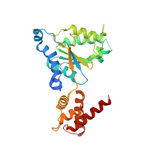
Structural basis for proteasome formation controlled by an assembly chaperone nas2.

Satoh, T.Saeki, Y.Hiromoto, T.Wang, Y.H.Uekusa, Y.Yagi, H.Yoshihara, H.Yagi-Utsumi, M.Mizushima, T.Tanaka, K.Kato, K.

(2014) Structure 22: 731-743

  • DOI: https://doi.org/10.1016/j.str.2014.02.014
  • Primary Citation of Related Structures:  
    3WHJ, 3WHK, 3WHL

  • PubMed Abstract: 

    Proteasome formation does not occur due to spontaneous self-organization but results from a highly ordered process assisted by several assembly chaperones. The assembly of the proteasome ATPase subunits is assisted by four client-specific chaperones, of which three have been structurally resolved. Here, we provide the structural basis for the working mechanisms of the last, hereto structurally uncharacterized assembly chaperone, Nas2. We revealed that Nas2 binds to the Rpt5 subunit in a bivalent mode: the N-terminal helical domain of Nas2 masks the Rpt1-interacting surface of Rpt5, whereas its C-terminal PDZ domain caps the C-terminal proteasome-activating motif. Thus, Nas2 operates as a proteasome activation blocker, offering a checkpoint during the formation of the 19S ATPase prior to its docking onto the proteolytic 20S core particle.


  • Organizational Affiliation

    Graduate School of Pharmaceutical Sciences, Nagoya City University, 3-1 Tanabe-dori, Mizuho-ku, Nagoya 467-8603, Japan; JST, PRESTO, 3-1 Tanabe-dori, Mizuho-ku, Nagoya 467-8603, Japan.


Macromolecules
Find similar proteins by:  (by identity cutoff)  |  3D Structure
Entity ID: 1
MoleculeChains Sequence LengthOrganismDetailsImage
Proteasome-activating nucleotidase, 26S protease regulatory subunit 6A
A, B, C, D, E
A, B, C, D, E, F, G, H
270Pyrococcus furiosus DSM 3638Saccharomyces cerevisiae S288C
This entity is chimeric
Mutation(s): 0 
Gene Names: panPF0115O3258RPT5YOR117WYOR3258WYTA1
UniProt
Find proteins for Q8U4H3 (Pyrococcus furiosus (strain ATCC 43587 / DSM 3638 / JCM 8422 / Vc1))
Explore Q8U4H3 
Go to UniProtKB:  Q8U4H3
Find proteins for P33297 (Saccharomyces cerevisiae (strain ATCC 204508 / S288c))
Explore P33297 
Go to UniProtKB:  P33297
Entity Groups  
Sequence Clusters30% Identity50% Identity70% Identity90% Identity95% Identity100% Identity
UniProt GroupsP33297Q8U4H3
Sequence Annotations
Expand
  • Reference Sequence
Experimental Data & Validation

Experimental Data

  • Method: X-RAY DIFFRACTION
  • Resolution: 2.60 Å
  • R-Value Free: 0.275 
  • R-Value Work: 0.242 
  • R-Value Observed: 0.243 
  • Space Group: P 1
Unit Cell:
Length ( Å )Angle ( ˚ )
a = 71.04α = 90.04
b = 85.76β = 90.03
c = 105.26γ = 89.89
Software Package:
Software NamePurpose
HKL-2000data collection
MOLREPphasing
REFMACrefinement
DENZOdata reduction
SCALEPACKdata scaling

Structure Validation

View Full Validation Report


Ligand Structure Quality Assessment 


Entry History 

Deposition Data

Revision History  (Full details and data files)

  • Version 1.0: 2014-03-26
    Type: Initial release
  • Version 1.1: 2014-04-16
    Changes: Database references
  • Version 1.2: 2017-08-16
    Changes: Refinement description, Source and taxonomy
  • Version 1.3: 2022-08-24
    Changes: Database references, Derived calculations
  • Version 1.4: 2023-11-08
    Changes: Data collection, Refinement description