3WEC

Structure of P450 RauA (CYP1050A1) complexed with a biosynthetic intermediate of aurachin RE


Experimental Data Snapshot

  • Method: X-RAY DIFFRACTION
  • Resolution: 2.19 Å
  • R-Value Free: 0.263 
  • R-Value Work: 0.214 
  • R-Value Observed: 0.216 

wwPDB Validation   3D Report Full Report


Ligand Structure Quality Assessment 


This is version 1.1 of the entry. See complete history


Literature

Structure of the quinoline N-hydroxylating cytochrome P450 RauA, an essential enzyme that confers antibiotic activity on aurachin alkaloids

Yasutake, Y.Kitagawa, W.Hata, M.Nishioka, T.Ozaki, T.Nishiyama, M.Kuzuyama, T.Tamura, T.

(2014) FEBS Lett 588: 105-110

  • DOI: https://doi.org/10.1016/j.febslet.2013.11.016
  • Primary Citation of Related Structures:  
    3WEC

  • PubMed Abstract: 

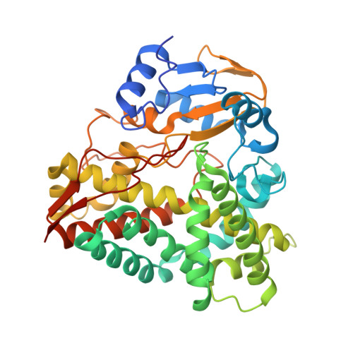
    The cytochrome P450 RauA from Rhodococcus erythropolis JCM 6824 catalyzes the hydroxylation of a nitrogen atom in the quinolone ring of aurachin, thereby conferring strong antibiotic activity on the aurachin alkaloid. Here, we report the crystal structure of RauA in complex with its substrate, a biosynthetic intermediate of aurachin RE. Clear electron density showed that the quinolone ring is oriented parallel to the porphyrin plane of the heme cofactor, while the farnesyl chain curls into a U-shape topology and is buried inside the solvent-inaccessible hydrophobic interior of RauA. The nearest atom from the heme iron is the quinolone nitrogen (4.3Å), which is consistent with RauA catalyzing the N-hydroxylation of the quinolone ring to produce mature aurachin RE.


  • Organizational Affiliation

    Bioproduction Research Institute, National Institute of Advanced Industrial Science and Technology (AIST), 2-17-2-1 Tsukisamu-Higashi, Toyohira-ku, Sapporo 062-8517, Japan. Electronic address: y-yasutake@aist.go.jp.


Macromolecules
Find similar proteins by:  (by identity cutoff)  |  3D Structure
Entity ID: 1
MoleculeChains Sequence LengthOrganismDetailsImage
Cytochrome P450419Rhodococcus erythropolisMutation(s): 0 
Gene Names: rauA
UniProt
Find proteins for S6BVH1 (Rhodococcus erythropolis)
Explore S6BVH1 
Go to UniProtKB:  S6BVH1
Entity Groups  
Sequence Clusters30% Identity50% Identity70% Identity90% Identity95% Identity100% Identity
UniProt GroupS6BVH1
Sequence Annotations
Expand
  • Reference Sequence
Small Molecules
Ligands 2 Unique
IDChains Name / Formula / InChI Key2D Diagram3D Interactions
HEM
Query on HEM

Download Ideal Coordinates CCD File 
B [auth A]PROTOPORPHYRIN IX CONTAINING FE
C34 H32 Fe N4 O4
KABFMIBPWCXCRK-RGGAHWMASA-L
AUI
Query on AUI

Download Ideal Coordinates CCD File 
C [auth A]3-[(2E,6E,9R)-9-hydroxy-3,7,11-trimethyldodeca-2,6,10-trien-1-yl]-2-methylquinolin-4(1H)-one
C25 H33 N O2
ODRSQPIWTIGZOH-ZDQFAWOQSA-N
Experimental Data & Validation

Experimental Data

  • Method: X-RAY DIFFRACTION
  • Resolution: 2.19 Å
  • R-Value Free: 0.263 
  • R-Value Work: 0.214 
  • R-Value Observed: 0.216 
  • Space Group: P 1 21 1
Unit Cell:
Length ( Å )Angle ( ˚ )
a = 41.46α = 90
b = 100.04β = 108.72
c = 52.29γ = 90
Software Package:
Software NamePurpose
ADSCdata collection
PHASERphasing
REFMACrefinement
MOSFLMdata reduction
SCALAdata scaling

Structure Validation

View Full Validation Report


Ligand Structure Quality Assessment 


Entry History 

Deposition Data

Revision History  (Full details and data files)

  • Version 1.0: 2014-01-01
    Type: Initial release
  • Version 1.1: 2023-11-08
    Changes: Data collection, Database references, Derived calculations, Refinement description