3WD6

Crystal structure of Bombyx mori omega-class glutathione transferase in complex with GSH


Experimental Data Snapshot

  • Method: X-RAY DIFFRACTION
  • Resolution: 2.50 Å
  • R-Value Free: 0.256 
  • R-Value Work: 0.208 
  • R-Value Observed: 0.210 

wwPDB Validation   3D Report Full Report


Ligand Structure Quality Assessment 


This is version 1.1 of the entry. See complete history


Literature

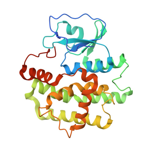
Three-dimensional structure of a Bombyx mori Omega-class glutathione transferase.

Yamamoto, K.Suzuki, M.Higashiura, A.Nakagawa, A.

(2013) Biochem Biophys Res Commun 438: 588-593

  • DOI: https://doi.org/10.1016/j.bbrc.2013.08.011
  • Primary Citation of Related Structures:  
    3WD6

  • PubMed Abstract: 

    Glutathione transferases (GSTs) are major phase II detoxification enzymes that play central roles in the defense against various environmental toxicants as well as oxidative stress. Here we report the crystal structure of an Omega-class glutathione transferase of Bombyx mori, bmGSTO, to gain insight into its catalytic mechanism. The structure of bmGSTO complexed with glutathione determined at a resolution of 2.5Å reveals that it exists as a dimer and is structurally similar to Omega-class GSTs with respect to its secondary and tertiary structures. Analysis of a complex between bmGSTO and glutathione showed that bound glutathione was localized to the glutathione-binding site (G-site). Site-directed mutagenesis of bmGSTO mutants indicated that amino acid residues Leu62, Lys65, Lys77, Val78, Glu91 and Ser92 in the G-site contribute to catalytic activity.


  • Organizational Affiliation

    Faculty of Agriculture, Kyushu University Graduate School, 6-10-1 Hakozaki, Higashi-ku, Fukuoka 812-8581, Japan. yamamok@agr.kyushu-u.ac.jp


Macromolecules
Find similar proteins by:  (by identity cutoff)  |  3D Structure
Entity ID: 1
MoleculeChains Sequence LengthOrganismDetailsImage
Omega-class glutathione S-transferase
A, B, C, D
256Bombyx moriMutation(s): 0 
Gene Names: gstoGSTo2
UniProt
Find proteins for A8R5V3 (Bombyx mori)
Explore A8R5V3 
Go to UniProtKB:  A8R5V3
Entity Groups  
Sequence Clusters30% Identity50% Identity70% Identity90% Identity95% Identity100% Identity
UniProt GroupA8R5V3
Sequence Annotations
Expand
  • Reference Sequence
Small Molecules
Ligands 5 Unique
IDChains Name / Formula / InChI Key2D Diagram3D Interactions
GSH
Query on GSH

Download Ideal Coordinates CCD File 
J [auth A],
N [auth B],
T [auth C],
X [auth D]
GLUTATHIONE
C10 H17 N3 O6 S
RWSXRVCMGQZWBV-WDSKDSINSA-N
IOD
Query on IOD

Download Ideal Coordinates CCD File 
O [auth C],
U [auth D]
IODIDE ION
I
XMBWDFGMSWQBCA-UHFFFAOYSA-M
PEG
Query on PEG

Download Ideal Coordinates CCD File 
I [auth A]DI(HYDROXYETHYL)ETHER
C4 H10 O3
MTHSVFCYNBDYFN-UHFFFAOYSA-N
EDO
Query on EDO

Download Ideal Coordinates CCD File 
H [auth A],
M [auth B],
R [auth C],
S [auth C],
W [auth D]
1,2-ETHANEDIOL
C2 H6 O2
LYCAIKOWRPUZTN-UHFFFAOYSA-N
K
Query on K

Download Ideal Coordinates CCD File 
E [auth A]
F [auth A]
G [auth A]
K [auth B]
L [auth B]
E [auth A],
F [auth A],
G [auth A],
K [auth B],
L [auth B],
P [auth C],
Q [auth C],
V [auth D]
POTASSIUM ION
K
NPYPAHLBTDXSSS-UHFFFAOYSA-N
Experimental Data & Validation

Experimental Data

  • Method: X-RAY DIFFRACTION
  • Resolution: 2.50 Å
  • R-Value Free: 0.256 
  • R-Value Work: 0.208 
  • R-Value Observed: 0.210 
  • Space Group: P 2 21 21
Unit Cell:
Length ( Å )Angle ( ˚ )
a = 75.861α = 90
b = 89.894β = 90
c = 182.149γ = 90
Software Package:
Software NamePurpose
HKL-2000data collection
MERLOTphasing
PHENIXrefinement
HKL-2000data reduction
HKL-2000data scaling

Structure Validation

View Full Validation Report


Ligand Structure Quality Assessment 


Entry History 

Deposition Data

Revision History  (Full details and data files)

  • Version 1.0: 2014-07-16
    Type: Initial release
  • Version 1.1: 2024-03-20
    Changes: Data collection, Database references, Derived calculations