


Structural insights into crRNA G-rich sequence binding and R-loop formation facilitated by Meiothermus ruber CasB
Yuan, Y.A., Yuan, Z.To be published.
Experimental Data Snapshot
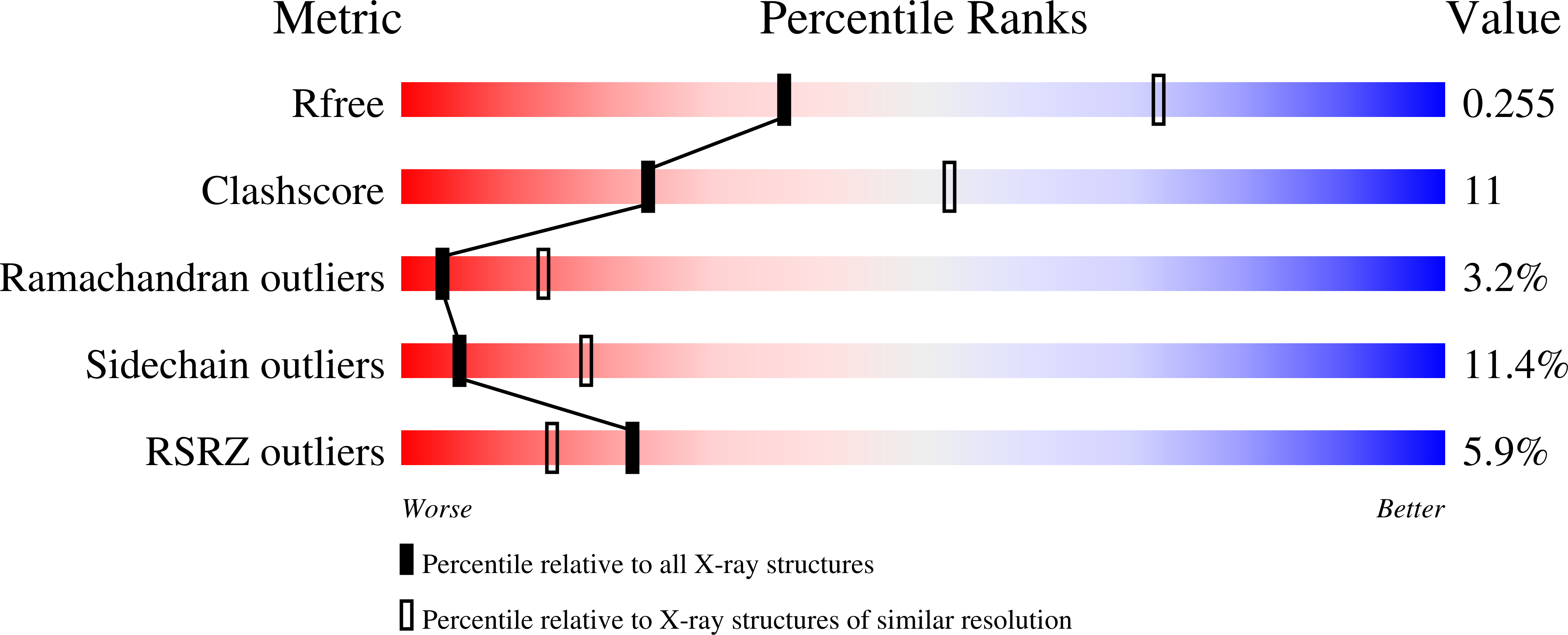
wwPDB Validation 3D Report Full Report
Entity ID: 1 | |||||
|---|---|---|---|---|---|
| Molecule | Chains | Sequence Length | Organism | Details | Image |
| CRISPR-associated protein, Cse2 family | 205 | Meiothermus ruber DSM 1279 | Mutation(s): 0 Gene Names: Mrub_3018 |  | |
UniProt | |||||
Find proteins for D3PQC9 (Meiothermus ruber (strain ATCC 35948 / DSM 1279 / VKM B-1258 / 21)) Explore D3PQC9 Go to UniProtKB: D3PQC9 | |||||
Entity Groups | |||||
| Sequence Clusters | 30% Identity50% Identity70% Identity90% Identity95% Identity100% Identity | ||||
| UniProt Group | D3PQC9 | ||||
Sequence AnnotationsExpand | |||||
| |||||
| Ligands 1 Unique | |||||
|---|---|---|---|---|---|
| ID | Chains | Name / Formula / InChI Key | 2D Diagram | 3D Interactions | |
| HG Query on HG | C [auth A], D [auth A], E [auth B], F [auth B] | MERCURY (II) ION Hg BQPIGGFYSBELGY-UHFFFAOYSA-N |  | ||
| Length ( Å ) | Angle ( ˚ ) |
|---|---|
| a = 71.823 | α = 90 |
| b = 75.383 | β = 90 |
| c = 112.671 | γ = 90 |
| Software Name | Purpose |
|---|---|
| HKL-2000 | data collection |
| SHARP | phasing |
| REFMAC | refinement |
| HKL-2000 | data reduction |
| HKL-2000 | data scaling |
RCSB PDB (citation) is hosted by
RCSB PDB is a member of the

