3W9Y

Crystal structure of the human DLG1 guanylate kinase domain


Experimental Data Snapshot

  • Method: X-RAY DIFFRACTION
  • Resolution: 2.20 Å
  • R-Value Free: 0.265 
  • R-Value Work: 0.209 
  • R-Value Observed: 0.215 

wwPDB Validation   3D Report Full Report


This is version 1.2 of the entry. See complete history


Literature

Crystal structure of the guanylate kinase domain from discs large homolog 1 (DLG1/SAP97)

Mori, S.Tezuka, Y.Arakawa, A.Handa, N.Shirouzu, M.Akiyama, T.Yokoyama, S.

(2013) Biochem Biophys Res Commun 435: 334-338

  • DOI: https://doi.org/10.1016/j.bbrc.2013.04.056
  • Primary Citation of Related Structures:  
    3W9Y

  • PubMed Abstract: 

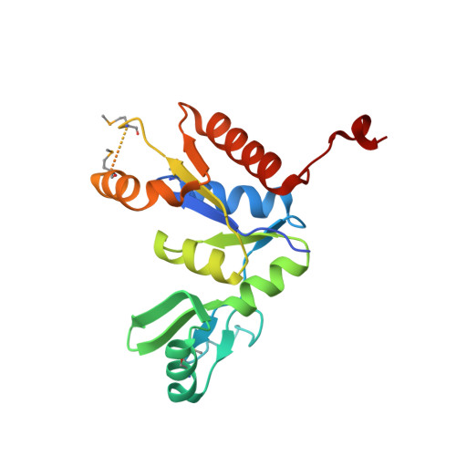
    Discs large homolog 1 (DLG1/SAP97) is involved in the development and regulation of neuronal and immunological synapses. DLG1 is a member of the membrane associated guanylate kinase (MAGUK) family of proteins, which function as molecular scaffolds. The C-terminal guanylate kinase (GK) domain of DLG1 binds peptides with a phosphorylated serine residue. In this study, we solved the crystal structure of the GK domain of human DLG1. The C-terminal tail of DLG1 is bound to the peptide-binding site of an adjacent symmetry-related DLG1 GK molecule. The binding direction of the C-terminal tail to the peptide-binding site is opposite to that of the phosphorylated LGN peptide in complex with the rat DLG1 GK domain. The C-terminal tail forms a 310 helix, which is also different from the conformation of the phosphorylated LGN peptide. Nevertheless, the side chain interactions of the C-terminal tail with the DLG1 GK domain are similar to those of the phosphorylated LGN peptide.


  • Organizational Affiliation

    Department of Biophysics and Biochemistry, Graduate School of Science, The University of Tokyo, Tokyo 113-0033, Japan.


Macromolecules
Find similar proteins by:  (by identity cutoff)  |  3D Structure
Entity ID: 1
MoleculeChains Sequence LengthOrganismDetailsImage
Disks large homolog 1200Homo sapiensMutation(s): 0 
Gene Names: DLG1
UniProt & NIH Common Fund Data Resources
Find proteins for Q12959 (Homo sapiens)
Explore Q12959 
Go to UniProtKB:  Q12959
PHAROS:  Q12959
GTEx:  ENSG00000075711 
Entity Groups  
Sequence Clusters30% Identity50% Identity70% Identity90% Identity95% Identity100% Identity
UniProt GroupQ12959
Sequence Annotations
Expand
  • Reference Sequence
Small Molecules
Modified Residues  1 Unique
IDChains TypeFormula2D DiagramParent
MSE
Query on MSE
A
L-PEPTIDE LINKINGC5 H11 N O2 SeMET
Experimental Data & Validation

Experimental Data

  • Method: X-RAY DIFFRACTION
  • Resolution: 2.20 Å
  • R-Value Free: 0.265 
  • R-Value Work: 0.209 
  • R-Value Observed: 0.215 
  • Space Group: P 65 2 2
Unit Cell:
Length ( Å )Angle ( ˚ )
a = 65.535α = 90
b = 65.535β = 90
c = 180.563γ = 120
Software Package:
Software NamePurpose
HKL-2000data collection
PHASERphasing
PHENIXrefinement
CNSrefinement
HKL-2000data reduction
HKL-2000data scaling

Structure Validation

View Full Validation Report


Entry History 

Deposition Data

Revision History  (Full details and data files)

  • Version 1.0: 2013-06-26
    Type: Initial release
  • Version 1.1: 2023-11-08
    Changes: Data collection, Database references, Derived calculations, Refinement description
  • Version 1.2: 2023-12-06
    Changes: Data collection