3W32

EGFR kinase domain complexed with compound 20a


Experimental Data Snapshot

  • Method: X-RAY DIFFRACTION
  • Resolution: 1.80 Å
  • R-Value Free: 0.236 
  • R-Value Work: 0.200 
  • R-Value Observed: 0.202 

wwPDB Validation   3D Report Full Report


Ligand Structure Quality Assessment 


This is version 1.2 of the entry. See complete history


Literature

Design and synthesis of novel pyrimido[4,5-b]azepine derivatives as HER2/EGFR dual inhibitors

Kawakita, Y.Seto, M.Ohashi, T.Tamura, T.Yusa, T.Miki, H.Iwata, H.Kamiguchi, H.Tanaka, T.Sogabe, S.Ohta, Y.Ishikawa, T.

(2013) Bioorg Med Chem 21: 2250-2261

  • DOI: https://doi.org/10.1016/j.bmc.2013.02.014
  • Primary Citation of Related Structures:  
    3W32, 3W33

  • PubMed Abstract: 

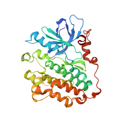
    A novel 7,6 fused bicyclic scaffold, pyrimido[4,5-b]azepine was designed to fit into the ATP binding site of the HER2/EGFR proteins. The synthesis of this scaffold was accomplished by an intramolecular Claisen-type condensation. As the results of optimization lead us to 4-anilino and 6-functional groups, we discovered 6-substituted amide derivative 19b, which has a 1-benzothiophen-4-yloxy group attached to the 4-anilino group. An X-ray co-crystal structure of 19b with EGFR demonstrated that the N-1 and N-3 nitrogens of the pyrimido[4,5-b]azepine scaffold make hydrogen-bonding interactions with the main chain NH of Met793 and the side chain of Thr854 via a water-mediated hydrogen bond network, respectively. In addition, the NH proton at the 9-position makes an additional hydrogen bond with the carbonyl group of Met793, as we expected. Compound 19b revealed potent HER2/EGFR kinase (IC50: 24/36 nM) and BT474 cell growth (GI50: 18 nM) inhibitory activities based on its pseudo-irreversible (PI) profile.


  • Organizational Affiliation

    Pharmaceutical Research Division, Takeda Pharmaceutical Co., Ltd, 26-1, Muraoka-Higashi 2-chome, Fujisawa, Kanagawa 251-8555, Japan. Electronic address: youichi.kawakita@takeda.com.


Macromolecules
Find similar proteins by:  (by identity cutoff)  |  3D Structure
Entity ID: 1
MoleculeChains Sequence LengthOrganismDetailsImage
Epidermal growth factor receptor330Homo sapiensMutation(s): 0 
Gene Names: EGFRERBBERBB1HER1
EC: 2.7.10.1
UniProt & NIH Common Fund Data Resources
Find proteins for P00533 (Homo sapiens)
Explore P00533 
Go to UniProtKB:  P00533
PHAROS:  P00533
GTEx:  ENSG00000146648 
Entity Groups  
Sequence Clusters30% Identity50% Identity70% Identity90% Identity95% Identity100% Identity
UniProt GroupP00533
Sequence Annotations
Expand
  • Reference Sequence
Small Molecules
Ligands 2 Unique
IDChains Name / Formula / InChI Key2D Diagram3D Interactions
W32
Query on W32

Download Ideal Coordinates CCD File 
B [auth A]4-({3-chloro-4-[3-(trifluoromethyl)phenoxy]phenyl}amino)-N-[2-(methylsulfonyl)ethyl]-8,9-dihydro-7H-pyrimido[4,5-b]azepine-6-carboxamide
C25 H23 Cl F3 N5 O4 S
QRXMEGUJRZKMEO-UHFFFAOYSA-N
SO4
Query on SO4

Download Ideal Coordinates CCD File 
C [auth A]SULFATE ION
O4 S
QAOWNCQODCNURD-UHFFFAOYSA-L
Binding Affinity Annotations 
IDSourceBinding Affinity
W32 Binding MOAD:  3W32 IC50: 75 (nM) from 1 assay(s)
BindingDB:  3W32 IC50: 75 (nM) from 1 assay(s)
PDBBind:  3W32 IC50: 75 (nM) from 1 assay(s)
Experimental Data & Validation

Experimental Data

  • Method: X-RAY DIFFRACTION
  • Resolution: 1.80 Å
  • R-Value Free: 0.236 
  • R-Value Work: 0.200 
  • R-Value Observed: 0.202 
  • Space Group: P 21 21 21
Unit Cell:
Length ( Å )Angle ( ˚ )
a = 46.443α = 90
b = 68.306β = 90
c = 103.273γ = 90
Software Package:
Software NamePurpose
ADSCdata collection
MOLREPphasing
REFMACrefinement
HKL-2000data reduction
HKL-2000data scaling

Structure Validation

View Full Validation Report


Ligand Structure Quality Assessment 


Entry History 

Deposition Data

Revision History  (Full details and data files)

  • Version 1.0: 2013-03-06
    Type: Initial release
  • Version 1.1: 2013-08-14
    Changes: Database references
  • Version 1.2: 2023-11-08
    Changes: Data collection, Database references, Derived calculations, Refinement description