3VZA

Crystal structure of the chicken Spc24-Spc25 globular domain in complex with CENP-T peptide


Experimental Data Snapshot

  • Method: X-RAY DIFFRACTION
  • Resolution: 1.90 Å
  • R-Value Free: 0.216 
  • R-Value Work: 0.167 
  • R-Value Observed: 0.170 

wwPDB Validation   3D Report Full Report


This is version 1.1 of the entry. See complete history


Literature

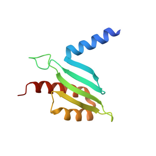
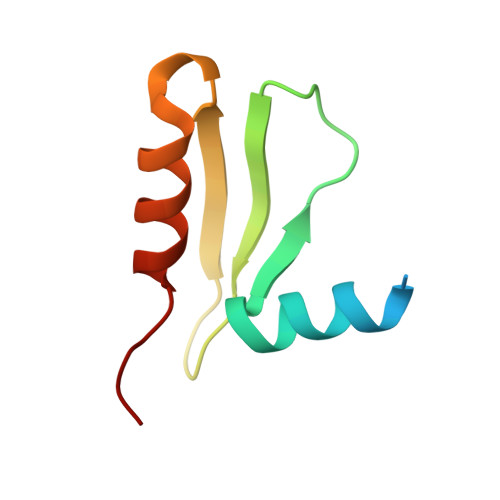
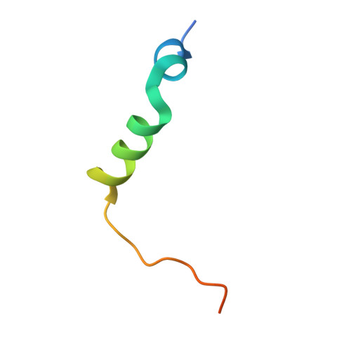
CENP-T provides a structural platform for outer kinetochore assembly

Nishino, T.Rago, F.Hori, T.Tomii, K.Cheeseman, I.M.Fukagawa, T.

(2013) EMBO J 32: 424-436

  • DOI: https://doi.org/10.1038/emboj.2012.348
  • Primary Citation of Related Structures:  
    3VZ9, 3VZA

  • PubMed Abstract: 

    The kinetochore forms a dynamic interface with microtubules from the mitotic spindle during mitosis. The Ndc80 complex acts as the key microtubule-binding complex at kinetochores. However, it is unclear how the Ndc80 complex associates with the inner kinetochore proteins that assemble upon centromeric chromatin. Here, based on a high-resolution structural analysis, we demonstrate that the N-terminal region of vertebrate CENP-T interacts with the 'RWD' domain in the Spc24/25 portion of the Ndc80 complex. Phosphorylation of CENP-T strengthens a cryptic hydrophobic interaction between CENP-T and Spc25 resulting in a phospho-regulated interaction that occurs without direct recognition of the phosphorylated residue. The Ndc80 complex interacts with both CENP-T and the Mis12 complex, but we find that these interactions are mutually exclusive, supporting a model in which two distinct pathways target the Ndc80 complex to kinetochores. Our results provide a model for how the multiple protein complexes at kinetochores associate in a phospho-regulated manner.


  • Organizational Affiliation

    Department of Molecular Genetics, National Institute of Genetics and The Graduate University for Advanced Studies SOKENDAI, Shizuoka 411-8540, Japan.


Macromolecules
Find similar proteins by:  (by identity cutoff)  |  3D Structure
Entity ID: 1
MoleculeChains Sequence LengthOrganismDetailsImage
Uncharacterized proteinA [auth B],
C [auth A]
105Gallus gallusMutation(s): 0 
Gene Names: SPC25
Entity Groups  
Sequence Clusters30% Identity50% Identity70% Identity90% Identity95% Identity100% Identity
Sequence Annotations
Expand
  • Reference Sequence
Find similar proteins by:  (by identity cutoff)  |  3D Structure
Entity ID: 2
MoleculeChains Sequence LengthOrganismDetailsImage
Spc24 proteinB [auth D],
D [auth C]
73Gallus gallusMutation(s): 0 
UniProt
Find proteins for R4GRT4 (Gallus gallus)
Explore R4GRT4 
Go to UniProtKB:  R4GRT4
Entity Groups  
Sequence Clusters30% Identity50% Identity70% Identity90% Identity95% Identity100% Identity
UniProt GroupR4GRT4
Sequence Annotations
Expand
  • Reference Sequence
Find similar proteins by:  (by identity cutoff)  |  3D Structure
Entity ID: 3
MoleculeChains Sequence LengthOrganismDetailsImage
Centromere protein T
E, F
39Gallus gallusMutation(s): 2 
Gene Names: CENPT
UniProt
Find proteins for F1NPG5 (Gallus gallus)
Explore F1NPG5 
Go to UniProtKB:  F1NPG5
Entity Groups  
Sequence Clusters30% Identity50% Identity70% Identity90% Identity95% Identity100% Identity
UniProt GroupF1NPG5
Sequence Annotations
Expand
  • Reference Sequence
Experimental Data & Validation

Experimental Data

  • Method: X-RAY DIFFRACTION
  • Resolution: 1.90 Å
  • R-Value Free: 0.216 
  • R-Value Work: 0.167 
  • R-Value Observed: 0.170 
  • Space Group: P 31
Unit Cell:
Length ( Å )Angle ( ˚ )
a = 61.321α = 90
b = 61.321β = 90
c = 111.275γ = 120
Software Package:
Software NamePurpose
HKL-2000data collection
PHENIXmodel building
PHENIXrefinement
HKL-2000data reduction
HKL-2000data scaling
PHENIXphasing

Structure Validation

View Full Validation Report


Entry History 

Deposition Data

Revision History  (Full details and data files)

  • Version 1.0: 2013-04-03
    Type: Initial release
  • Version 1.1: 2023-11-08
    Changes: Data collection, Database references, Refinement description