3VTA

Crystal Structure of cucumisin, a subtilisin-like endoprotease from Cucumis melo L


Experimental Data Snapshot

  • Method: X-RAY DIFFRACTION
  • Resolution: 2.75 Å
  • R-Value Free: 0.262 
  • R-Value Work: 0.214 
  • R-Value Observed: 0.214 

wwPDB Validation   3D Report Full Report


Ligand Structure Quality Assessment 


This is version 2.0 of the entry. See complete history


Literature

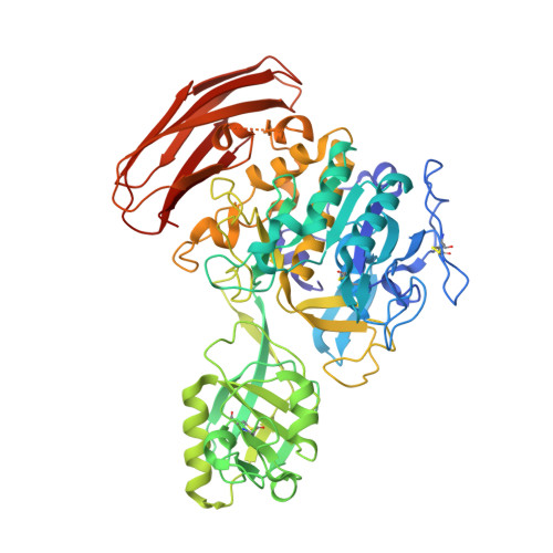
Crystal structure of cucumisin, a subtilisin-like endoprotease from Cucumis melo L

Murayama, K.Kato-Murayama, M.Hosaka, T.Sotokawauchi, A.Yokoyama, S.Arima, K.Shirouzu, M.

(2012) J Mol Biol 423: 386-396

  • DOI: https://doi.org/10.1016/j.jmb.2012.07.013
  • Primary Citation of Related Structures:  
    3VTA

  • PubMed Abstract: 

    Cucumisin is a plant serine protease, isolated as an extracellular glycoprotein from the melon fruit Cucumis melo L. var. Prince. Cucumisin is composed of multiple domain modules, including catalytic, protease-associated, and fibronectin-III-like domains. The crystal structure of cucumisin was determined by the multiwavelength anomalous dispersion method and refined at 2.75Å resolution. A structural homology search indicated that the catalytic domain of cucumisin shares structural similarity with subtilisin and subtilisin-like fold enzymes. According to the Z-score, the highest structural similarity is with tomato subtilase 3 (SBT3), with an rmsd of 3.5Å for the entire region. The dimer formation mediated by the protease-associated domain in SBT3 is a distinctive structural characteristic of cucumisin. On the other hand, analytical ultracentrifugation indicated that cucumisin is mainly monomeric in solution. Although the locations of the amino acid residues composing the catalytic triad are well conserved between cucumisin and SBT3, a disulfide bond is uniquely located near the active site of cucumisin. The steric circumstances of the active site with this disulfide bond are distinct from those of SBT3, and it contributes to the substrate preference of cucumisin, especially at the P2 position. Among the plant serine proteases, the thermostability of cucumisin is higher than that of its structural homologue SBT3, as determined by their melting points. A structural comparison between cucumisin and SBT3 revealed that cucumisin possesses less surface area and shortened loop regions. Consequently, the higher thermostability of cucumisin is achieved by its more compact structure.


  • Organizational Affiliation

    Division of Biomedical Measurements and Diagnostics, Graduate School of Biomedical Engineering, Tohoku University, Sendai 980-8575, Japan.


Macromolecules
Find similar proteins by:  (by identity cutoff)  |  3D Structure
Entity ID: 1
MoleculeChains Sequence LengthOrganismDetailsImage
Cucumisin
A, B
621Cucumis meloMutation(s): 0 
EC: 3.4.21.25
UniProt
Find proteins for Q39547 (Cucumis melo)
Explore Q39547 
Go to UniProtKB:  Q39547
Entity Groups  
Sequence Clusters30% Identity50% Identity70% Identity90% Identity95% Identity100% Identity
UniProt GroupQ39547
Sequence Annotations
Expand
  • Reference Sequence
Oligosaccharides

Help

Entity ID: 2
MoleculeChains Length2D Diagram Glycosylation3D Interactions
alpha-D-mannopyranose-(1-6)-beta-D-mannopyranose-(1-4)-2-acetamido-2-deoxy-beta-D-glucopyranose-(1-4)-[alpha-L-fucopyranose-(1-3)]2-acetamido-2-deoxy-beta-D-glucopyranose
C, D
5N-Glycosylation
Glycosylation Resources
GlyTouCan:  G83878UZ
GlyCosmos:  G83878UZ
GlyGen:  G83878UZ
Entity ID: 3
MoleculeChains Length2D Diagram Glycosylation3D Interactions
alpha-L-fucopyranose-(1-3)-[2-acetamido-2-deoxy-beta-D-glucopyranose-(1-4)]2-acetamido-2-deoxy-beta-D-glucopyranose
E
3N-Glycosylation
Glycosylation Resources
GlyTouCan:  G11971MR
GlyCosmos:  G11971MR
GlyGen:  G11971MR
Small Molecules
Experimental Data & Validation

Experimental Data

  • Method: X-RAY DIFFRACTION
  • Resolution: 2.75 Å
  • R-Value Free: 0.262 
  • R-Value Work: 0.214 
  • R-Value Observed: 0.214 
  • Space Group: H 3
Unit Cell:
Length ( Å )Angle ( ˚ )
a = 149.483α = 90
b = 149.483β = 90
c = 218.035γ = 120
Software Package:
Software NamePurpose
HKL-2000data collection
SOLVEphasing
CNSrefinement
HKL-2000data reduction
HKL-2000data scaling

Structure Validation

View Full Validation Report


Ligand Structure Quality Assessment 


Entry History 

Deposition Data

Revision History  (Full details and data files)

  • Version 1.0: 2012-08-08
    Type: Initial release
  • Version 1.1: 2013-07-24
    Changes: Database references
  • Version 2.0: 2020-07-29
    Type: Remediation
    Reason: Carbohydrate remediation
    Changes: Atomic model, Data collection, Derived calculations, Structure summary