3VRM

Structure of cytochrome P450 Vdh mutant T107A with bound vitamin D3


Experimental Data Snapshot

  • Method: X-RAY DIFFRACTION
  • Resolution: 2.57 Å
  • R-Value Free: 0.231 
  • R-Value Work: 0.187 
  • R-Value Observed: 0.189 

wwPDB Validation   3D Report Full Report


Ligand Structure Quality Assessment 


This is version 1.2 of the entry. See complete history


Literature

A single mutation at the ferredoxin binding site of p450 vdh enables efficient biocatalytic production of 25-hydroxyvitamin d3.

Yasutake, Y.Nishioka, T.Imoto, N.Tamura, T.

(2013) Chembiochem 14: 2284-2291

  • DOI: https://doi.org/10.1002/cbic.201300386
  • Primary Citation of Related Structures:  
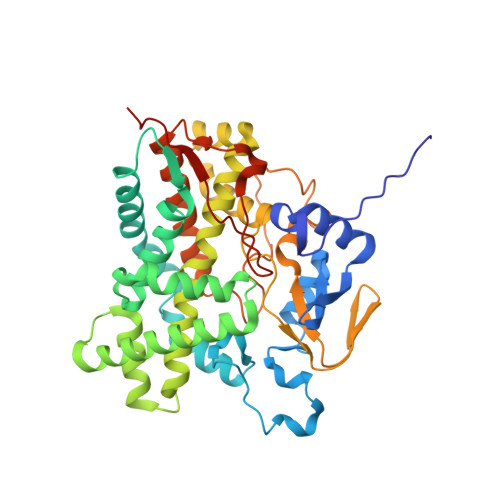
    3VRM

  • PubMed Abstract: 

    Vitamin D3 hydroxylase (Vdh) from Pseudonocardia autotrophica is a cytochrome P450 monooxygenase that catalyzes the two-step hydroxylation of vitamin D3 (VD3 ) to produce 25-hydroxyvitamin D3 (25(OH)VD3 ) and 1α,25-dihydroxyvitamin D3 (1α,25(OH)2 VD3 ). These hydroxylated forms of VD3 are useful as pharmaceuticals for the treatment of conditions associated with VD3 deficiency and VD3 metabolic disorder. Herein, we describe the creation of a highly active T107A mutant of Vdh by engineering the putative ferredoxin-binding site. Crystallographic and kinetic analyses indicate that the T107A mutation results in conformational change from an open to a closed state, thereby increasing the binding affinity with ferredoxin. We also report the efficient biocatalytic synthesis of 25(OH)VD3 , a promising intermediate for the synthesis of various hydroxylated VD3 derivatives, by using nisin-treated Rhodococcus erythropolis cells containing VdhT107A . The gene-expression cassette encoding Bacillus megaterium glucose dehydrogenase-IV was inserted into the R. erythropolis chromosome and expressed to avoid exhaustion of NADH in a cytoplasm during bioconversion. As a result, approximately 573 μg mL(-1) 25(OH)VD3 was successfully produced by a 2 h bioconversion.


  • Organizational Affiliation

    Bioproduction Research Institute, National Institute of Advanced Industrial Science and Technology (AIST), 2-17-2-1, Tsukisamu-higashi, Toyohira-ku, Sapporo 062-8517 (Japan).


Macromolecules
Find similar proteins by:  (by identity cutoff)  |  3D Structure
Entity ID: 1
MoleculeChains Sequence LengthOrganismDetailsImage
Vitamin D(3) 25-hydroxylase411Pseudonocardia autotrophicaMutation(s): 1 
Gene Names: vdh
EC: 1.14.13.15
UniProt
Find proteins for C4B644 (Pseudonocardia autotrophica)
Explore C4B644 
Go to UniProtKB:  C4B644
Entity Groups  
Sequence Clusters30% Identity50% Identity70% Identity90% Identity95% Identity100% Identity
UniProt GroupC4B644
Sequence Annotations
Expand
  • Reference Sequence
Small Molecules
Ligands 2 Unique
IDChains Name / Formula / InChI Key2D Diagram3D Interactions
HEM
Query on HEM

Download Ideal Coordinates CCD File 
B [auth A]PROTOPORPHYRIN IX CONTAINING FE
C34 H32 Fe N4 O4
KABFMIBPWCXCRK-RGGAHWMASA-L
VD3
Query on VD3

Download Ideal Coordinates CCD File 
C [auth A](1S,3Z)-3-[(2E)-2-[(1R,3AR,7AS)-7A-METHYL-1-[(2R)-6-METHYLHEPTAN-2-YL]-2,3,3A,5,6,7-HEXAHYDRO-1H-INDEN-4-YLIDENE]ETHYLI DENE]-4-METHYLIDENE-CYCLOHEXAN-1-OL
C27 H44 O
QYSXJUFSXHHAJI-RWDMXNMGSA-N
Experimental Data & Validation

Experimental Data

  • Method: X-RAY DIFFRACTION
  • Resolution: 2.57 Å
  • R-Value Free: 0.231 
  • R-Value Work: 0.187 
  • R-Value Observed: 0.189 
  • Space Group: C 2 2 21
Unit Cell:
Length ( Å )Angle ( ˚ )
a = 61.421α = 90
b = 105.521β = 90
c = 141.988γ = 90
Software Package:
Software NamePurpose
ADSCdata collection
MOLREPphasing
REFMACrefinement
XDSdata reduction
XDSdata scaling

Structure Validation

View Full Validation Report


Ligand Structure Quality Assessment 


Entry History 

Deposition Data

Revision History  (Full details and data files)

  • Version 1.0: 2013-04-17
    Type: Initial release
  • Version 1.1: 2013-11-27
    Changes: Database references
  • Version 1.2: 2023-11-08
    Changes: Data collection, Database references, Derived calculations, Refinement description, Structure summary